Prepared by the researche : Majida Hameed Obaida1 and Saif Jabbar Yasir2
- Al-Furat Al-Awsat Technical University, Najaf Technical Institute, AL Najaf, Iraq.
- Department of Medical Microbiology, College of Medicine, University of Kufa, Najaf, Iraq
DAC Democratic Arabic Center GmbH
Journal of Progressive Medical Sciences : Second issue – August 2025
A Periodical International Journal published by the “Democratic Arab Center” Germany – Berlin
ISSN 3052-8518
Journal of Progressive Medical Sciences
:To download the pdf version of the research papers, please visit the following link
Abstract
Earth’s most common viruses, bacteriophages, infect bacteria and archaea. Protein capsids and sometimes lipid envelopes encase phage genomes, which vary in kind and structure. Most are double-stranded DNA phages, whereas Cystoviridae are RNA. Phages control bacterial populations, transfer horizontal genes, lyse molecules, and alter metabolism in different situations. Based on their structural morphotypes, which are largely determined by tail architecture, phages are classified as Podoviridae (short tails), Siphoviridae (long, flexible tails), and Myoviridae (long, contractile tails). They build genome transport and host recognition virions with portal proteins, tail fibers, and baseplates. DNA is transferred into capsids by ATP-dependent terminases during genome packing. The lysogenic cycle comprises temperate phages integrating into the host genome as prophages and replicating passively until stimulation activates the lytic phase. Phages with non-lytic chronic infections or carriers exist. Phages limit their host range by recognizing lipopolysaccharides, outer membrane proteins, teichoic acids, and flagella. They can make anti-CRISPR proteins to attack bacteria. Phages indirectly affect human health by influencing microbiota and immune systems. Immunoglobulin-like capsid domains connect them to mucosal surfaces for bacterial clearance and barrier protection. They can also enter tissues and circulation, where they are immunologically tolerated. Phages activate innate and adaptive immunity. Innate sensing and cytokine production are enabled by TLR3, TLR7, and TLR9. However, adaptive immunity produces neutralizing IgM, IgG, and IgA antibodies. Recurrent phage therapy can be reduced by phage-neutralizing antibodies. Phage biology is characterized by the discovery of “huge phages” with genomes that rival small bacterial genomes and encode complex functions like tRNAs, translation factors, CRISPR-Cas systems, and nucleus-like compartments that protect phage genomes from host defenses. Phage-encoded CRISPR-Cas systems often lack spacer acquisition or interference genes, resulting in host apparatus repurposing or gene transcriptional silence. Targeting competing phages and host regulatory mechanisms is possible. Phages treat antibiotic-resistant microorganisms. Phage treatment has potential despite bacterial resistance and host immune neutralization. They also carry toxin genes (cholera, diphtheria) through lysogenic conversion, boosting bacterial pathogenicity. Dynamic evolutionary arms races affect microbial ecology through phage-host and phage-phage interactions. Recent metagenomics and meta transcriptomics advances have increased the variety of dsRNA and tailless dsDNA phages. This showed unique viral families and the prevalence of phages in human microbiomes, particularly crAss-like phages in the intestine. The molecular details of phage interactions with eukaryotic cells are still emerging, despite the vast knowledge of bacterial receptors for phage attachment. Mammalian cells can use endocytosis mechanisms to internalize phages for immunological regulation and therapeutic delivery. expanding the taxonomy of dsRNA phages, understanding non-lytic infection mechanisms, characterizing phage-host range and interactions, and using phages for biocontrol in agriculture and medicine. It is required to overcome immune clearance, understand phage immunogenicity, and understand the tri-kingdom interactions between phages, bacteria, and human hosts that maintain microbial and immunological homeostasis to maximize phage therapy.
Bacteriophages are complex, diversified, and ecologically important viruses. Bacteria, horizontal gene transfer, immune system contact, and burgeoning biological uses including phage therapy and biotechnology are managed. Phages’ complex life cycles, structural biology, and interactions with human and bacterial hosts are being understood beyond their role as bacterial predators. Briefly explain bacteriophages, which infect bacteria and archaea, their diversity, dsDNA and dsRNA phages, structure and replication mechanisms, ecological implications, mammalian immunity, and emerging therapeutic applications, including phage therapy. Phage viruses are assembled by complex structural proteins. Widely studied tailed phages including T7 (Autographviridae/Podoviridae-like), SPP1 (Siphoviridae-like), and T4 (Myoviridae-like) have tail and capsid shapes optimized for genome transport and host recognition Biomolecules such lipopolysaccharides and outer membrane proteins bind to baseplate and tail fibers. Genome packing motor ATPases assist phage DNA enter capsids. T4 phages employ the lytic cycle to rapidly multiply and lyse the host cell, while temperate phages use the lysogenic cycle to passively integrate their genome into the host chromosome until induction activates lytic replication. Infection-causing pseudolysogenic or persistent phages do not lyse quickly. In bacterial pathogen phage treatment, these life choices affect phage ecology and application. To identify hosts, phages recognize polysaccharides, proteins, and flagella. Phage host range is limited by specificity. To escape bacterial defenses, phages can encode anti-CRISPR proteins and adapt genomically. Phages transfer toxin genes and other virulence factors to bacteria by lysogenic conversion. Phages influence microbiomes and immunity without infecting humans. Phages can infect tissues and cells via endocytosis, altering immune responses. Phages destroy bacteria to fight antibiotic-resistant illnesses, but immunological responses including neutralizing antibodies can hinder phage treatment. Recent discoveries include “huge phages” with microscopic bacteria genomes. Complex machinery is provided by tRNA, translation factor, and CRISPR-Cas phages. Metagenomic studies have found tailless dsDNA phages from new viral orders and families, enhancing our understanding of phage diversity. CRISPR-mediated phage-host and phage-phage interactions show dynamic evolutionary arms races. Biomedical applications, gene transfer, microbial community dynamics, and bacterial population regulation depend on bacteriophages. Research on their structural biology, life cycles, and host immunological interactions determines their therapeutic and ecological potential. Bacteriophages are everywhere in the biosphere and influence bacterial ecology and evolution. DNA-containing bacteriophages in ecosystems are well-studied, whereas RNA-containing ones are often overlooked.
Classification and Diversity:
The ICTV has classified more than 50 virus families of double-stranded DNA bacteriophages. The Cystoviridae is the sole family of dsRNA bacteriophages, consisting of seven species. In contrast to dsDNA phages, classified dsRNA phage isolates exhibit a restricted host range. Most phages are double-stranded DNA tailed (order Caudovirales), but double-stranded RNA (family Cystoviridae) and tailless phages also contribute to viral variety. New metagenomic research have revealed a great diversity of phages, including giant phages with massive genomes that encode complicated functionalities including tRNAs, translation factors, and CRISPR-Cas systems. Phylogenetic investigations of signature genes like RNA-dependent RNA polymerase and capsid protein structures are currently used more than morphology or host specificity to progress taxonomy.
In the present taxonomy, dsRNA viruses are classified as either Pisuviricota or Duplornaviricota. The majority of animal dsRNA viruses (e.g., rotavirus and bluetongue virus) and several significant microbial and plant viruses are members of the Cystoviridae virus family (class: Vidaverviricetes, order: Mindivirales) of the phylum Duplornaviricota. The phylum Pisuviricota comprises dsRNA viruses, including amalga-, curvula-, partiti-, and picobirnaviruses. This initial taxonomy classification of RNA viruses is based on a comprehensive sequence-based comparison of viral RdRp sequences [1]. Nevertheless, a recent phylogenetic analysis of RdRp sequences based on sequences revealed that cystoviral and dsRNA virus RdRps are classified in Pisuviricota [2]. Additionally, higher-order groupings were identified through a structure-based phylogenetic analysis of viral RdRps [3].
The Cystoviridae family was recognized by the ICTV in 1978 [4], and the phi6 group and phage phi6 were recognized in 1976 [5]. At present, the Cystoviridae virus family comprises a single genus, Cystovirus (formerly known as the phi6 group), which comprises seven virus species: phi6, phi8, phi12, phi13, phiNN, phi2954, and phiYY. Due to their unique similarities in virion structures and genomes, these phages were classified together, despite their low nucleotide sequence identity (<50%, with the exception of phi6 and phiNN; [6]. The current criteria for identifying Cystoviridae species are 95% nucleotide sequence identity. The Cystoviridae family comprises six viruses that remain unidentified: phi7, phi9–phi11, phi14, and phiNY. phiZ98 and CAP3-7 may also be included in the Cystoviridae family due to their genetic and structural similarities [7,8]. The ICTV has recently introduced supplementary higher-order ranks for virus taxonomic classification and is in the process of transitioning to a system that more accurately reflects virus phylogenetic relationships than host specificity, morphological features, or disease symptoms [9]. The polymerase gene, which encodes RdRp or reverse transcriptase, is a critical locus for the classification of RNA viruses and phylogenetic analysis. RdRp-encoding RNA viruses are found in the Orthornavirae kingdom of the Riboviria domain. Duplornaviricota, Kitrinoviricota, Lenarviricota, Negarnaviricota, and Pisuviricota are the five official phyla of Orthornavirae [10].
In the recently adopted megataxonomy of viruses, double-stranded DNA phages are divided into two huge realms: Duplodnaviria and Varidnaviria [11]. Viral structures in these realms include MCP and packing ATPases. Archaeal, tailed, and herpesviruses are duplodnaviria. The phylum Nucleocytoviricota, which comprises mimiviruses, tailless bacteriophages, and related archaeal viruses, is included in Varidnaviria.
In cultivation, tailed Duplodnaviria bacteriophages are the most prevalent. The upper oceans are dominated by non-tailed varidnaviruses, which account for 50% to 90% of viral particles, according to metagenomic investigations [121,13]. Varidnaviria’s icosahedral capsids are composed of the double jelly-roll (DJR) MCP [14,15]. The DJR MCP core structure is conserved across the entire spectrum of Varidnaviria members that infect hosts from all three domains of life, despite the fact that some viruses are difficult to recognize and the sequences are highly varied [16,17]. By employing sensitive profile-based methods to search metagenomic sequence data for DJR MCP contigs, an unexpectedly extensive array of varidnaviruses was identified, which are likely to infect bacterial and archaeal hosts. This diversity surpassed that of prokaryote viruses in the families Turriviridae, Tectiviridae, Corticoviridae, and Autolykiviridae in the class Tectiliviricetes [18]. In the ‘PM2-like’ category, which included several prophages, tailless phages were most prevalent. This group includes two Corticoviridae phages, PM2 [19] and Cr39582 [20], eleven Vibrio phages of the Autolykiviridae family, and the unassigned f No16 phage [21], which infect Pseudoalteromonas species. Vinavirales includes Corticoviridae and Autolykiviridae [22]. Most phages in this group have 9-12 kb genomes that are circular (Corticoviridae) or have inverted terminal repeats. Vinavirales viruses encode structural components and genes that govern transcription, genome replication, and cell lysis. Proviruses from Vinavirales members are extensively incorporated into aquatic bacteria genomes. Their occurrence in other habitats is unknown. DJR MCP and packing ATPase are the only Vinavirales-shared proteins [23]. Metagenomics has been used to study the human intestine virome, a major microbiome factor. Most identified viruses are Duplodnaviria domain tailed bacteriophages [24,25,26]. The most common human-associated viruses are crAss-like phages, which the ICTV recently categorized into the Crassvirales order. These phages were discovered by metagenomics investigation and have a wide variety of very varied members. Gut metagenomics has found many new abundant-tailed bacteriophage families [27,28] Varidnaviria members are likely a minor component of the gastrointestinal virome and are not well-known. Study conducted a search of 23,119 gut metagenomes for DJR MCP and discovered that a significant number of the sequences encoding this hallmark protein of Varidnaviria are associated with prophages that are distantly related to the families Corticoviridae and Autolykiviridae. These prophages suggest a new Vinavirales order with at least three families. It found an extended, conserved gene core of Vinavirales with 12 genes, one of which encodes a hitherto undiscovered lysin protein, using sensitive protein structure prediction and sequence analysis methods. [29,30.31]
Structure and composition:
Unlike cells, phages viruses that infect bacteria cannot perform most biological processes needed for reproduction. They modify ecosystems by preying on certain bacterial populations, mediating lateral gene transfer, altering host metabolism, and redistributing bacterial chemicals by cell lysis [23,33,34,35,26]. They spread antibiotic resistance and pathogenic agents that infect humans and animals [37,38]. Our knowledge of phages comes from laboratory studies, most of which had genomes a few tens of kb. Many isolation methods remove big phage particles from phage concentrations obtained by 100-nm or 200-nm filters. Only 93 isolated phages with genomes over 200 kb were published in 2017 [32].
Community-wide DNA sequencing is capable of detecting phage-derived fragments; however, fragmentation can obscure large genomes. A novel clade of megaphages associated with humans and animals was identified in the genomes of metagenomic datasets that were manually curated [39]. In order to ascertain the prevalence, diversity, and environmental distribution of large-genome phages, we implemented a more comprehensive investigation of microbial communities. In the past, phages with genomes over 200 kb were called “jumbophages” or megaphages[40]. We call the set represented here “huge phages” because it spans both sizes. A graphical abstract summarizes our approach and findings. This work shows the wide range of environments where phages have genomes as large as small-celled bacteria. We hypothesize that these phages have evolved a unique “life” strategy that entails substantial host biology interception and enhancement while replicating their massive genomes. [41,42,43].[44] were able to reconstruct 351 phages, 6 plasmid-like, and 4 unclassified sequences. It exclusively retained CRISPR–Cas loci and excluded plasmids. We incorporated three phage sequences that were ≤200 kb in length as a result of the CRISPR-Cas loci. As anticipated, we identified numerous phage-relevant genes, such as those that are involved in the encoding of structural proteins and lysis, as well as other genomic properties of phages. It was anticipated that certain structural proteins would be as long as 7,694 amino acids. 175 phage sequences were circularized, and 35 were manually curated to completion, occasionally by resolving complex repetition sections to disclose encoded proteins. Although the majority of genomes are incomplete, a few may be complete but linear. Bidirectional replication is indicated by the GC asymmetry of 30% of genomes, while unidirectional replication is indicated by 30% of genomes [45]. The largest phage genomes known are our 4 largest complete, carefully curated, and circularized genomes, 634, 636, 642, and 735 kb. The largest circularized phage genome known was 596 kb [46]. The prior work reported a 630-kb circularized genome, but it was an assembly artifact. Concatenation artefacts were so severe in IMG/VR [47] that we excluded these data from future analyses. We revised our phage genome size distribution using full and circularized genomes from our study and published genomes. Without the enormous phages mentioned here, entire phages had median genomic sizes of 52 kb. Thus, these sequences greatly increase the number of phages with extremely large genomes. [44]
Nine of our genomes have coding densities that are less than 78%, which may be attributed to a genetic code that deviates from the standard code. This effect is uncommon in phages; however, it has been observed in Lak phage. The UAG stop codon appears to have been reassigned to encode an amino acid in numerous genomes, primarily human and animal. By transitioning into a flanking bacterial genome sequence, only one region exceeding 200 kb was identified as a prophage. Nevertheless, prophage integration is feasible, as half of the genomes were not circularized. Genomes containing integrases suggest a temperate lifestyle in certain circumstances. [48] There are significant structural differences within these groupings, especially in tail tips and capsid diameters. Since the tail tube is isolated, this knowledge is enough to place the main tail protein in its structural context. Capsid, fiber, and baseplate proteins will also be skipped.
SPP1 is a phage of the tail-morphotype that is similar to Siphoviridae. The dodecameric portal protein gp6 is located on one vertex of the icosahedral procapsid of the SPP1 phage. The maturation of procapsid into capsid is facilitated by the translocation of viral DNA into the head and the release of Gp6 [49,50]. ATP is utilized by the packaging terminase to facilitate the passage of DNA through the portal complex. The hexameric ring–shaped adaptor protein complex gp15 and stopper protein complex gp16 attach to the portal complex after capsid formation to limit DNA leakage, resulting in the formation of the head-to-tail connector [51]. The tail of SPP1 is dependent on the hexameric ring-shaped distal tail protein (Dit) gp19.1. The distal tail adsorption function and the tail tube [52] are connected by this junction. The length of the tail tube [53] is determined by 40 stacked hexameric rings of MTP gp17.1 and its C-terminally extended form gp17.1∗, which are constructed around a tape measure protein gp8, which may be trimeric. A ribosomal frameshift leads to the C-terminal expansion of gp17.1∗, which contains an immunoglobulin (Ig) fold and a fibronectin type III (FN3) domain. The tail tube is composed of gp17.1 and gp17.1∗ in a 3:1 ratio. Nevertheless, virions that contain only gp17.1 are infectious [54]. The FN3 domain may transiently interact with Bacillus subtilis cell wall carbohydrates, thereby increasing infection, during two-dimensional diffusion of virions on the outer bacterial surface. The tail tube top is tapered by a hexameric ring of the tail completion protein gp17, which binds to the stopper protein gp16 and connects the tail to the head-to-tail connector. The trimeric tail tip protein gp21, as well as potentially gp22, gp23, gp23.1, and gp24, are involved in the distal tail adsorption function [55]. The positions of these proteins are ambiguous. The YueB receptor ectodomain of Bacillus subtilis is irreversibly bound by gp21 or a protein, resulting in infection. The formation of the tail in λ phages commences with an initiator complex that includes tape measure protein, assembly chaperones, distal tail protein, baseplate proteins, and tail tip proteins. This combination enables MTP to polymerize onto the distal tail protein ring and tape measure protein, resulting in the formation of a helical tube that replaces assembly chaperones. When the tail tube completely engulfs the tape measure protein, MTP oligomerization concludes. The tail completion protein will truncate the tail and connect it to the head-to-tail connector to complete the virion [56].
In contrast, SPP1 phage MTP (gp17.1) can self-polymerize without protein [57]. The tail creation process may differ. Dissociating the tail tip after binding to the YueB receptor primes the release of the metastable tape measure protein from the tail tube. Next, the stopper protein gp16 opens diaphragm-like and ejects DNA into the tail tube to start infection [58]. Tape measure proteins may produce host membrane pores for DNA translocation [59 ]. T4 phage is a Myoviridae-like tail-morphotype phage. This phage has a more complicated head assembly than the others: The membrane-spanning initiation complex of the dodecameric portal protein gp20 and gp40 binds 11 scaffolding proteins to start procapsid assembly [60 ]. The prohead’s fivefold vertices in the capsid shell are produced by the major capsid protein gp23 and the vertex protein gp24. Procapsids are released from the membrane and space is created for DNA by proteolytic cleavage of the scaffolding and capsid proteins [61]. The ATP-dependent terminase translocates DNA through the portal complex and expands the procapsid into the final capsid. This process creates binding sites for the small outer capsid protein (gp soc) and the highly antigenic outer capsid protein (gp hoc) that decorate the capsid [62]. Icosahedral extremities and cylindrical equatorial central regions are features of the T4 phage capsid. The head is completed and the portal complex is sealed by the binding of the Gp13–gp14 neck complex. The six segments of the baseplate are arranged around a tube [63] to initiate the tail assembly.
The junction for tail tube polymerization [64] at the proximal end of the baseplate is composed of hexameric rings of gp48 and gp54, while the tail tip at the distal end is composed of gp27, gp5, and gp5.4. The trimeric proteins gp5 and gp5.4 puncture host membranes and hydrolyze host peptidoglycan, while gp27 is capable of passing DNA. To create the tail tube, the MTP gp19 polymerizes into 24 hexameric rings around the baseplate-anchored tape measure protein gp29 [65]. The tail tube terminator protein gp3 [66] taper the proximal end of the tail tube. Gp18, the sheath protein, undergoes helical polymerization around the tail tube [67]. Gp15 completes the tail by binding to gp3 and the terminal ring of the tail sheath. The virion is completed by the interaction between gp15 and (gp13–gp14), which connects the tail and head-to-tail connector. T4 is equipped with a head whisker at the head-to-tail connector and short and long tail fibers at the baseplate. The short tail fibers unwind from beneath the baseplate and irreversibly bond to LPS, anchoring the baseplate to the outer membrane, when E. coli’s long tail fibers bind to LPSs and OmpC of the outer membrane [68]. The tail sheath is constricted by the baseplate’s reorganizations, which compel the tail tip to pass through the outer membrane. This process digests the peptidoglycan layer and translocates the tape measure protein gp29 and viral DNA through the tail tube. An inner membrane pore may be generated by the tape measure protein gp29 and/or gp27, which enables DNA to enter the host cytoplasm [69]. Phage virions have complicated designs for host recognition, genome packaging, and genome delivery, with tail morphologies resembling Podoviridae, Siphoviridae, and Myoviridae families. Mechanisms of phage assembly and infection reveal coordinated protein interactions and ATP-driven DNA packaging motors. Unique RNA bacteriophages with lipid envelopes and multilayered capsids, enveloped dsRNA phages (Cystoviridae) share structural similarities with eukaryotic dsRNA viruses, suggesting evolutionary linkages.
Currently, phi6 and phi12 are the best-characterized dsRNA phages, their component proteins, and assembly intermediates. However, similar gene sets in known dsRNA phage isolates suggest a similar virion organization. However, new studies show structural variance, especially in host recognition and entrance components. Phi6, phi8, phi12, phi2954, phiNN, phiYY, phiNY, phiZ98, and CAP3 have enveloped spherical virions in negative-stain transmission electron microscopy (TEM) [70,71], cryo-EM, and cryo-ET [72]. Detergent or organic solvent sensitivity studies have shown that dsRNA phage virion lipids exist.
These are the sole membrane-enveloped RNA bacteriophages that are currently known. phi6 and phi8 virions exhibited envelope spikes of 2 and 7 nm following cryo-EM characterization. These structures are host attachment spikes produced by multimeric P3 complexes, as indicated by prior research on P3-deficient phi6 virions [73]. Subsequently, cryo-ET and three-dimensional reconstruction revealed toroidal or elongated structures on phi12 and phi2954 virions. By employing the S and L segments from phi12 and the M segment from phi2954, a recombinant phi12 phage was generated through reverse genetic technique. The recombinant phage exhibited host specificity and envelope surface features similar to those of phi2954 [74].
By dsRNA phage envelopes, icosahedrally symmetric nucleocapsids are enclosed. The nucleocapsid surface shell and polymerase complex were identified in these particles in the initial EM investigation on phi6 virions [75]. The protein P8 trimer nucleocapsid shell is arranged into an incomplete icosahedral T = 13 lattice, which is interrupted at the five-fold symmetry positions by P4 complexes protruding from the polymerase complex layer, as demonstrated by high-resolution cryo-EM imaging and three-dimensional reconstruction analyses of phages phi6 and phi12 [76]. The cystovirus phi8 deviates from this fundamental structure by lacking a nucleocapsid shell. Phi6 bonds to and penetrates the host plasma membrane through the P8 layer [77]. This suggests that polymerase complex components are responsible for these critical responsibilities, as purified phi8 core particles have the capacity to infect spheroplasts of host cells [78]. Additional research is necessary to ascertain the presence of a nucleocapsid surface shell that enables plasma membrane penetration in other dsRNA phages that are similar. The phi6 polymerase complex, which consists of the proteins P1, P2, P4, and P7, replicates and transcribes the viral dsRNA. Cryo-EM has identified dimers of the primary inner capsid protein (MCP) P1 on an icosahedral T = 1 lattice in the empty and/or genome-containing polymerase complex particles of phi6, phi8, and phi12 [79]. I find it intriguing that the capsid organization of picobirnaviruses, Reovirales order animal and plant dsRNA viruses, and members of the families Partitiviridae, Totiviridae, Chrysoviridae, Quadriviridae, and Megabirnaviridae is similar (referred to as “T = 2”) [80,81]. This organization is not present in any other viruses. This suggests that dsRNA viral capsids have a shared ancestry.
The P2 structure of the phi6 polymerase subunit was described by Butcher et al. in 2001. The first high-resolution structure of RNA-dependent RNA polymerase (RdRp) from dsRNA virus. The phi6 P2 structure was found to be remarkably similar to the RdRp of the hepatitis C virus, indicating that the polymerase subunit of a dsRNA phage and a eukaryotic positive-sense single-stranded RNA virus may have shared a common evolutionary origin. Several high-resolution structures for additional viral RdRps have been deposited in the protein data bank over the past two decades. A structure-based computational comparison has demonstrated that the phi6 and phi12 RdRps [82] are structurally similar to all currently structurally characterized viral RdRps [83].
Protein P4 is one of the most extensively studied cystovirus proteins in terms of its physical and functional characteristics. P4, a molecular motor that organizes viral single-stranded genomic precursor molecules into empty polymerase complexes, is powered by NTP hydrolysis. The structures of the P4 proteins of phi6, phi8, phi12, phi13, and phiYY are of high resolution [84]. Despite structural differences, these proteins form hexameric complexes that resemble RecA-type ATPases. A genome with three dsRNA segments: L (large, 6.3–7.1 kb), M (medium, 3.6–4.7 kb), and S is present in each Cystoviridae family member and related dsRNA phage isolate. The genome is in the range of 12.7–15.0 kb (phi2954–phi8). Comparative genomic analyses indicate that certain dsRNA phage isolates are closely associated with Pseudomonas phage phi6, while others are distant. The L, M, and S segments of Pseudomonas phages phiNN and phi6 exhibit 80%, 55%, and 84% nucleotide sequence identity, respectively. However, phiNY does not appear to share any similarities with other cystoviruses or phages.
The genome organizations of the six dsRNA phage isolates CAP3–7 and phiZ98, as well as the currently recognized cystoviruses, are comparable. The L segment encodes proteins for the polymerase complex (MCP P1, RdRp P2, packaging NTPase P4, and assembly factor P7), the M segment encodes proteins for host recognition and outer membrane penetration (P3 and P6), and the S segment encodes the nucleocapsid shell protein (P8), major membrane protein (P9), putative membrane morphogenetic factor (non-structural protein P12), and the RNA polymerase. In contrast, the majority of the open reading frames that are expected to be generated by phiNY contain proteins of unknown function, and only the RdRp and MCP genes in the L segment and the glycoside hydrolase gene in the S segment could be predicted, necessitating further investigation. Nevertheless, the coding regions are flanked by non-coding sections in all dsRNA phage genome segments. According to phi6 research, these non-coding regions are essential for genome packaging and replication. [85,86]. Despite their high gene synteny, certain cystoviruses possess open reading frames with ambiguous functions. The P3 host recognition spike complex contains a single P3 protein or its multimer in the spike complex of cystoviruses phi6, phi2954, and phiNN [87 ]. In contrast, the heteromeric spike complex of phi8, phi12, phi13, and phiYY contains two or three viral proteins [88]. Recent metatranscriptomic surveys indicate that dsRNA phages have developed a variety of lytic enzymes. Cystoviridae members encode lytic transglycosylases from the lysozyme superfamily. However, some cystovirus-like contigs encode putative N-acetylmuramoyl-L-alanine amidase, metallopeptidase (families M15 and M23), lipase, and L-alanyl-D-glutamate endopeptidase genes. The variety of lysis genes in these potential dsRNA phages may suggest a broader host range. [89]
Phage Replication Cycles and Infection Strategies:
Bacteriophages undergo lytic or lysogenous cycles. Furthermore, certain phages are pseudolysogenic. Proteins that are anticipated to locate the bacterial membrane or cell surface are encoded by the phage genomes. These factors may influence the susceptibility of the host to phage infection. Almost all of the host metabolism-enhancing genes that were previously identified were discovered. Numerous phages possess genes that synthesize purines and pyrimidines de novo and convert nucleic and ribonucleic acids and nucleotide phosphorylation states. These gene sets are reminiscent of microorganisms that are remarkably minuscule and likely have symbiotic relationships. Numerous phages contain transcription and translation genes. The unique sequences of up to 67 tRNAs are encoded by complete phage genomes. In general, the number of tRNAs increases as the genome length increases (Spearman’s ρ = 0.61, P = 4.5 × 10⁻²², n = 201). Each genome of large phages contains up to 15 tRNA synthetases that are distinct but related to those of their hosts. Via these proteins, phages can charge their tRNA variations with host amino acids. Genes for tRNA modification and ligation of host defense-cleaved tRNAs are present in certain genomes.
Genes that intercept and redirect host translation are present in numerous phages. Among these genes are the ribosomal proteins S4, S1, S21, and L7/L12, as well as the initiation factors IF1 and IF3. Ribosomes were recently discovered in phages. rpS1 and rpS21 are indispensable for the initiation of translation in bacteria, which renders them advantageous for the hijacking of host ribosomes. Additional investigation of rpS21 proteins revealed N-terminal extensions that contain RNA-binding basic and aromatic residues. The anticipation of that these phage ribosomal proteins will supplant host proteins and promote competitive ribosome binding or preferential phage mRNA initiation. Lytic phages, such as T4, lyse and eradicate bacterial cells following virion multiplication. Phage offspring are capable of infecting other organisms following the cell’s death. Lytic phages are more effective for therapeutic purposes. Some lytic phages prevent phage progeny from lysing out of the cell when extracellular phage concentrations are high.
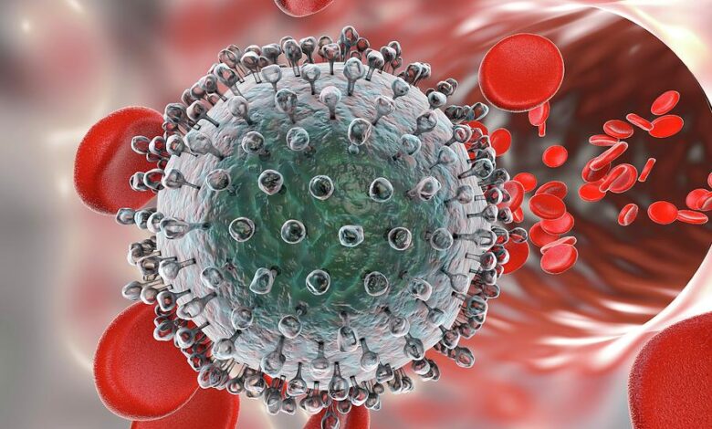
This process differs from temperate phage dormancy and is usually transient. [90] The lysogenic cycle does not immediately lyse the host cell. Lysogeny-capable phages are temperate. Their viral genome integrates with host DNA and replicates harmlessly or becomes a plasmid. Endogenous phages (prophages) activate when host circumstances decrease, such as nutritional deprivation. The host cell lyses when they start reproducing. The lysogenic cycle permits the host cell to live and proliferate, replicating the virus in all offspring. E. coli’s lambda phage follows the lysogenic and lytic cycles.[91] Prophages can help the host bacterium by adding additional functionalities to the genome during dormancy, called lysogenic conversion. Bacteriophages can turn safe strains of Corynebacterium diphtheriae or Vibrio cholerae into highly virulent ones that cause diphtheria or cholera. [92] There are ways to fight certain bacterial infections by targeting toxin-encoding prophages. [93] The viruses in this electron micrograph of bacteriophages adhering to a bacterial cell are coliphage-sized. T1
Bacterial cells’ polysaccharide cell walls shield them against antibiotics and immunological host defenses. [94] Bacteriophages bind to lipopolysaccharides, teichoic acids, proteins, or flagella on bacteria to enter a host cell. Bacteriophages can only infect bacteria with receptors they can bind to, limiting their host range. Virion-associated polysaccharide-degrading enzymes breakdown their hosts’ capsular outer coat during the start of a tightly regulated phage infection. Host growth parameters affect phage attachment and invasion. Phage virions cannot move autonomously, thus they must randomly contact the right receptors in solution, such as blood, lymphatic circulation, irrigation, soil water, etc. Myovirus bacteriophages inject genetic material into cells using a hypodermic syringe. The tail fibers flex to bring the base plate closer to the cell after hitting the receptor. This is reversible binding. Once fully joined, irreversible binding begins and the tail contracts, presumably with ATP, shooting genetic material through the bacterial membrane. [95] The shaft bends to the side, contracts closer to the cell, and pushes up to inject. Unlike myoviruses, podoviruses use their short, tooth-like tail fibers to enzymatically destroy a section of the cell membrane before introducing their genetic material.
In minutes, bacterial ribosomes convert viral mRNA into protein. Early RNA replicase synthesis occurs in RNA-based phages. Proteins make bacterial RNA polymerase prefer viral mRNA. The host must produce viral products instead of proteins and nucleic acids due to disruptions. Ribosomal proteins in some dsDNA bacteriophages may influence protein translation during infection. [96] T4 phage morphogenesis requires catalytic helper proteins to build new virus particles. The foundation plates are produced first, then the tails. Separately built head capsids will spontaneously assemble with tails. Phage genes encode morphogenetic proteins that interact in a specific sequence during phage T4 virion formation. Normal phage T4 morphogenesis requires a balance in these proteins produced during viral infection. DNA is efficiently packed in heads. The entire process takes 15 minutes.
studies of bacteriophage T4 (1962–1964) revealed almost all of the genes required for laboratory development. [97] Two groups of conditional lethal mutations enabled these experiments. Amber mutants were one type. Temperature-sensitive mutants were another type of conditional lethal mutant. These two groups of mutants revealed the activities and connections of proteins involved in DNA replication, repair, and recombination and how viruses are built from protein and nucleic acid components. Sometimes phages are released via cell lysis, extrusion, or budding. Tailed phages lyse by breaking down cell wall peptidoglycan with endolysin. Filamentous phages cause host cells to release new virus particles. Free virions can infect a new bacterium unless faulty. Budding is linked to Mycoplasma phages. Unlike virion release, lysogenic phages become prophages and stay in the host. [98]
The majority of cystovirus isolates are virulent and lyse the bacterial cells of their host during reproduction. Certain dsRNA phages have the ability to produce viral particles within the host cell without causing lysis or integrating into the host chromosome. This single-cell phenomenon be referred to as a non-productive chronic infection or carrier cell in order to differentiate it from the carrier state life cycle, which should be allocated to population-level phage-host interactions. In cell cultures that are continuously evolving, cystovirus carrier cells are identified by intracellular viral genomic dsRNA molecules or viral particles. phi6 and its P. syringae host were first reported to infect this way, but the phi13 variant (containing a kanamycin resistance gene) has also been found to infect Salmonella typhimurium and E. coli. Recently, dsRNA phage phiNY was shown to infect similarly. The continuous phiNY infection boosted Microvirgula aerodenitrificans host growth, suggesting a mutualistic, parasitic lifestyle. Interestingly, dsRNA phages persist like fungus dsRNA viruses, which do not create external infectious virions and are mostly cryptic.
Bacteriophages are essential to the human microbiome and mediate genetic exchange between pathogenic and nonpathogenic bacteria, even though they cannot infect and multiply in human cells. Transduction occurs when a bacteriophage transfers genes from one bacterial strain to another. Endocytosis pathways are key to eukaryotic cell bacteriophage uptake. Phages cannot multiply in eukaryotic cells but can be absorbed through cellular processes similar to those utilized for other particles or viruses: Phages can be taken up by clathrin-mediated, caveolin-dependent, or macropinocytosis from eukaryotic cells. Phages can enter vesicles and pass epithelial barriers via these internalization mechanisms [99]. Receptor Binding: Polysialic acid-binding E. coli phages can enter neuroblastoma cells by interacting with cell surface chemicals like polysialic acids [100]. After endocytosis, some phages can cross human epithelial cells to reach underlying tissues via intracellular trafficking processes that converge on recycling endosomes [99]. Phage Display and Cell Penetration Peptides: Engineered phages with TAT (transactivator of transcription) peptides increase membrane translocation and phage entry into various eukaryotic cells [101]. Phage uptake by eukaryotic cells is researched, revealing different methods by which phages can interact with and penetrate human cells, modulating immune responses and providing therapeutic benefits.
Recent experiments have illuminated how eukaryotic cells ingest bacteriophages: A 2023 study found that mammalian cells ingest bacteriophage T4 mostly by macropinocytosis, which engulfs extracellular fluid and particles. Intact phages accumulate in macropinosomes and go through endosomal pathways such lysosomal destruction or exocytosis. Importantly, internalized phages induced protein phosphorylation cascades that promoted cell metabolism and survival without activating inflammatory DNA-sensing immune pathways Phages promote cellular development and metabolism, which has implications for phage therapy and microbiome interactions [102]. Another experiment evaluated neuroblastoma cell internalization of an Escherichia coli bacteriophage (PK1A2) that binds to polysialic acid. Endolysosomal internalization of the phage-receptor complex was shown by fluorescence and electron microscopy. Phages remained intracellular for 24 hours. Phages were redistributed to vesicular structures surrounding the nucleus, confirming receptor-mediated endocytosis as a significant uptake mechanism [103]. Filamentous M13 phages with cell-penetrating peptides like TAT were shown to increase endocytosis and intracellular trafficking in mammalian cells. Phage surface changes promoted cellular penetration and therapeutic administration in this experiment [103]. Active cellular mechanisms such macropinocytosis and receptor-mediated endocytosis are key to human cell phage uptake, according to this research. They show endosomal phage trafficking and putative cellular signaling responses from internalized phages.
Some of the molecular receptors for bacteriophage binding on eukaryotic cells have been experimentally characterized: Polysilic acid: A well-known receptor on human neuroblastoma cells that binds and internalizes Escherichia coli phages. This sialic acid polymer aids receptor-mediated phage endocytosis into eukaryotic cells [105]. Sugars and glycans: Though less well-defined than bacterial phage receptors, eukaryotic cell surface carbohydrate moieties can bind phages [106]. Peptides that penetrate cells: Engineered peptides on phages like TAT interact with negatively charged membrane components to increase mammalian cell uptake, suggesting membrane phospholipid or proteoglycan components may help bind [107]. Many eukaryotic phage receptors’ molecular identities are unknown. In contrast, bacterial phage receptors typically contain outer membrane proteins, lipopolysaccharides, teichoic acids, and polysaccharides that have been extensively studied in numerous phage-host systems [108]. Thus, polysialic acid and certain surface glycans are major bacteriophage receptors for eukaryotic cells, while cell-penetrating peptide-targeted membrane components offer tailored uptake options.
Lytic cycles with fast host cell lysis or lysogenic cycles integrating their genome into host chromosomes allow persistence and horizontal gene transfer. Some dsRNA phages cause chronic non-lytic host cell infections. Tail fibers or spikes that identify lipopolysaccharides or proteins bind to bacterial cells specifically. Phage infection involves anti-CRISPR proteins and nucleus-like compartments that protect phage DNA to avoid host resistance.
Horizontal Gene Transfer, Phages:
Phages transfer horizontal genes by universal and specialized transduction, affecting bacterial virulence, antibiotic resistance, and evolution. Lysogenic conversion via prophages encodes toxins and other virulence factors, causing bacterial disease.
Transduction:
As the host cell disintegrates from lytic replication, generalized transduction bundles random bacterial genomic DNA in phage capsids instead of phage genomic DNA. The bacterium’s genome and that of its progeny cells may be altered if this phage injects this bacterial DNA into a healthy host cell and integrates into the bacterium’s chromosome. When initiating a lytic replication cycle in specialized transduction, lysogenic phages that have been amplified in a population of bacteria may eliminate a portion of the bacterial DNA with their genome. Lysogens share an integration site, which is why all progeny phages transfer the same bacterial gene to their hosts.
In addition to genetic exchange, bacteriophages can affect microbial populations by preying on select bacteria species while ignoring others. This characteristic has been studied to treat pathogenic bacterial infections in humans and animals for over 100 years. Wild phages may temporarily affect wild bacterial populations, [109], but lytic bacteriophages as antimicrobial therapy (phage therapy) in humans face significant challenges. Various wild bacterial strains resist phages. Famous resistance mechanisms include the CRISPR-Cas9 system, which was created for genetic manipulation in the lab and started as a bacterial defensive mechanism against bacteriophage invasion. [110] Phages are more immunogenic than antimicrobials and removed from the blood by the reticular endothelial system quickly. If effective phage mixtures are developed, their size may limit their topical use compared to antimicrobials. Some researchers recommend using phage enzymes, which can permeate bacterial cell walls, for simplicity. [111] No randomized, controlled, double-blind trials have proved either technique works in humans.
Host Specificity and Phage-Host Interactions
Bacteriophage-human cell interactions are a new field of study that combines microbiology and immunology and reveals exciting intricacies beyond basic bacteria-phage dynamics. The gut is full of phages, viruses that infect bacteria, which regulate bacterial ecosystems and immunological activities. Phages can directly interact with human cells, affecting immunological responses, inflammation, and tissue homeostasis, according to recent studies. Phages seldom infect humans, although they can adhere to eukaryotic cells and be internalized, influencing cell signaling pathways and immunological activation.
Besides altering microbiota, phages interact with human cells in therapeutic ways. Phages can infiltrate human macrophages and epithelial cells and give antibiotic effects to intracellular microorganisms. This intracellular trip of phages implies a sophisticated way they can manage bacterial illnesses inaccessible to some drugs. Phages also interact with receptors on human immune cells to control innate immune responses and shape inflammatory and antiviral defenses. Understanding that phages’ interactions with human cells rely on the type of phage, the bacterial environment, and the host’s immunological condition is crucial. Some phages have developed unique tactics to avoid human immune detection or alter host cells to help their bacterial hosts survive. Phages are not human pathogens, but their close interactions with human cells can affect gut homeostasis, immunological tolerance, and dysbiotic inflammation. Phages are being studied for their antibacterial and cell-interacting medicinal potential. Phage-human cell interactions reveal a dynamic dialogue where phages are more than bacterial predators. Phages modulate the immune system and control intracellular pathogens in human cells, offering a viable therapeutic avenue. Phages are natural partners in our microbiota, promoting health through diverse cellular interactions. [12-18].Phages, or bacteriophages, infect bacteria, but recent study has shown their complicated connections with human cells. Phages are plentiful in the gut, skin, and respiratory system, where they maintain microbial balance and prevent bacterial infections. Phages can attach directly to human cell surfaces, entering cells and affecting biological activities, offering promising therapeutic and immune system regulation potential. Phages are internalized by mammalian cells, which is critical for phage-human cell contact. Several investigations have shown that certain phages can endocytose human epithelial and immunological cells. Phages can interact with intracellular components, regulate signaling pathways, and deliver genetic material. Phages’ intracellular presence challenges the idea that they work only extracellularly and shows they may regulate host immunity and inflammation.
Phages directly stimulate immune cells or interact with bacterial populations. They activate immune cell pattern recognition receptors, producing cytokines and modulating inflammation. Phages are interesting candidates for innovative therapies for bacterial infections and inflammatory illnesses due to their immunomodulatory and antibacterial properties. Phages are typically safe for humans, with little negative effects in clinical trials. Phages’ capacity to interact with human cells is being used to treat antibiotic-resistant intracellular bacterial infections. Phages can enter host cells to reach buried germs, improving therapy success in persistent infections. Phage-host cell interactions are being studied to increase delivery, reduce resistance, and boost immunomodulation while maintaining safety. Besides bacterial targeting, bacteriophages and human cells interact directly, modulate the immune system, and have therapeutic potential. This new knowledge calls into question phages’ role in human physiology and immunological homeostasis as well as antibacterial agents. [19-126]. Phages change the bacterial microbiome and affect human physiology and immunology without infecting cells. Endocytosis or receptors allow phages to enter tissues and cells, altering mucosal immunity and cell activity. Microbial ecology and immunological homeostasis are regulated by a complex tri-kingdom interaction network of bacteriophages, bacteria, and human cells.
Interactions with Mammalian Immunity:
Clinical aspects:
Phages are clinically significant for many reasons. First, bacteriophage genomes encode several highly pathogenic bacterial toxins, making the host bacterium only pathogenic when lysogenized. Vibrio cholerae, Corynebacterium diphtheriae, Clostridium botulinum, Clostridium difficile, and Shigella species produce cholera, diphtheria, botulinum neurotoxin, binary, and Shiga toxins, respectively. [127]. These bacteria are either harmless or nonpathogenic without phage-encoded toxins. Phages encode these poisons for unknown reasons. Botulinum toxin paralysis seems to have the opposite effect of cholera toxin, which causes watery diarrhea and helps the phage and host find their next victim. Second, bacteriophages enable horizontal gene transfer, including antibiotic resistance. They have been developed to incorporate genes into specific strains for clinical use, but this is still in testing. [128] A third therapeutically relevant component of bacteriophages is their usage as a biomarker for their host in complicated environmental samples. This usually indicates water feces pollution. The host is likely present if the phage is. To identify bacteria in mixed environmental samples, phages have been designed to produce luciferase when they infect their host. Bacteriophages may identify strains of the same bacterial species, making them clinically valuable even though they are generally replaced by newer technology. As humans are susceptible to many viruses, most bacteria have multiple bacteriophage pathogens. Not all strains of a species are phage-resistant. Infecting each strain routinely with a standardized panel of phages for that species reveals its sensitivity and resistance to each phage type. To distinguish S. aureus strains, phage typing used a standardized panel of bacteriophages common internationally. Phage typing was the standard epidemiological strain monitoring approach before molecular methods like multilocus sequence typing and pulsed-field gel electrophoresis. Finally, bacteriophages were the first virus discovered and contributed to several molecular biology breakthroughs. Bacteriophages proved that DNA transferred genetic information, set up gene control, and revealed the genetic code. [129]
Immune Responses to Phages in Humans
Phages greatly influence the immune system: Innate immunity: Phages activate pattern recognition receptors, producing cytokines and modulating immunity. Adaptive immunity: Strong humoral responses generate IgM, IgG, and mucosal IgA antibodies that neutralize phages and alter clearance and therapy. Cellular adaptive responses include T cell activation. Repetition of phage therapy reduces bioavailability due to host immunological neutralization by antibodies and complement. Phages can cause pro- or anti-inflammatory immune responses depending on context and purity. Phages can influence immune cells to help eliminate microorganisms synergistically or inhibit immunological processes, demonstrating diverse immunomodulatory actions.
Phages’ indirect impact on mammalian immunity:
Bacterial disease, ecology, and genetic evolution are all significantly influenced by phages. Consequently, phages indirectly influence host defense and immunological function. Bacterial virulence and human immunity are influenced by the proteins encoded by phages. Lysogenic phages encode proteins that enable their bacterial hosts to penetrate tissue barriers, which serve as the initial line of defense against infections. The temperate phage Φctx is renowned for its ability to parasitize Vibrio cholerae hrough the production of cholera toxin. Bacterial adhesion, colonization, tissue invasion, and biofilm formation are all facilitated by phage-derived virulence factors [130,131].
The immune-clearing phagocytes are either destroyed or discouraged by phage-encoded proteins. The phage-encoded chemotaxis inhibitory protein of Staphylococcus aureus binds to and inhibits neutrophil receptors for complement and formylated proteins, thereby safeguarding it from neutrophil-mediated mortality. Exotoxin [132], cytotoxicity, intracellular infection [133], and superantigen are all produced or delivered by other phage-encoded proteins. Chromosome transcription factors primarily regulate phage-encoded virulence genes, which are synthesized during lysogeny, when other genes are not actively transcribed. For example, the lytic phage genes are located on the coding strand, while the λ-encoded bor of E. coli and the phage-encoded vir of M. arthritidis are located on the noncoding strand. Lastly, phages horizontally transfer antibiotic resistance genes within and between bacteria [134]. Prophages improve their bacterial hosts’ fitness and propagation. Prophages can shed genes, including virion-producing genes, and domesticate [135]. When they still benefit, prophage-derived genomic elements can be selectively retained [136]. Thus, many phage-bacteria interactions are complex coevolutionary interactions.
Direct Interactions with Phages and Innate Immunity:
Innate immunity controls the microbial-human contact through structural and germline-encoded characteristics. Phages may be essential to our relationship with bacterial flora, as they contribute to and cross these barriers. In the mucosa, circulation, and cells, phages are identified on the cell surface and in endocytic vesicles. Phages are abundant at bacterial colonization sites and may directly contribute to mucosal barrier defenses. Barr et al. [137] discovered that mucosal surfaces can retain as many as 109 adhering phages per biopsy. Phages can be modified to facilitate these interactions. Immunoglobulin (Ig)-like domains are present in E. coli T4 phage capsid proteins, which interact with epithelial cell mucins and surface glycoproteins. Many phage families include Ig superfamily-like protein domains, suggesting mucosal layer enrichment of other phages [138]. Mucosal binding increased the susceptibility of some bacteria to phage-mediated lysis [139] and phage diffusion in the mucus layer [140]. Thus, mucosal phages may prevent bacterial invasion in a ubiquitous, strain-specific, and non-host-derived manner. These findings suggest a complicated interaction between phages, bacteria, and mucosal surfaces that needs additional study. Antiphage antibodies may weaken or change barrier immunity, among other questions.
In mice models of colitis, the intestinal virome affects innate immunity, which may affect outcomes. Yang et al. [141] found that antiviral medicines worsened DSS-induced colitis in mice, but gut-resident viruses detected by TLR 3 and TLR7 protected via IFN-β production. Phages make up most of the intestinal virome, however this study did not focus on them. In contrast, Gogokhia et al. [142] found that oral phage cocktail exacerbated DSS colitis via TLR9. Phages’ significance in intestinal homeostasis needs further study. Mucosal-associated phages may affect tissues beyond the epithelial barrier. Transcytosis across cells transports many phages across the gut and into systemic circulation, possibly for paracytosis at inflammatory sites. Based on in vitro studies of cell monolayers [142], this transit is expected to occur at 3.1 × 10⁷ particles/day. Transcytosis across the Golgi apparatus allows phage T4 to absorb apically to basolaterally. Certain phages and peptide sequences may be more easily taken up [144]. T1 phage straight entering the small intestine entered gut-draining lymph and blood. Bioactivity against bacteria in circulating phages may help hosts defend against bloodstream bacteria. These investigations demonstrate the potential importance of phage interactions with mucosal immunity and how little we know about them. It is unclear how many phages transverse mucosal tissues, what processes are involved, if M cells, which sense the intestinal environment, aid transmission, or whether inflammation influences this transit.
Peripheral circulation and tissues contain several phages. The pharmacokinetics of some of these phages have been widely studied for lytic phage treatment. Regardless of method, circulating phages clear temporally and spatially. Phages last several days, with the biggest decline (>99%) in the first hour. After infusion, phages are discovered in most major organs, with the liver and spleen having the greatest and most lasting titers, indicating that these organs remove circulating phage particles. Splenic and hepatic macrophages phagocytose phage quickly and efficiently, according to research. Active phage is less persistent in the liver than in the spleen. Kupffer cells endocytose better, retain greater basal ROS levels, and produce fewer proinflammatory cytokines in response to TLR ligation than splenic macrophages [145]. These cells’ phagolysosomes digested and inactivated internalized phage. Phage clearance involves two steps: quick elimination of most particles in 24 hours, followed by slower clearance of the remaining ∼1% of circulating phages over many days. This slower tail may be phages trapped by antibodies or other immune response components. Inflammation and bacterial contamination may influence phage pharmacokinetics [146]. Immunologically, circulating and peripheral phages are well tolerated. Phages at immunological privilege areas including the placenta and CSF are also well tolerated. The recently obtained CSF virome shows that phage are abundant (104 pfu/mL) without inflammation [147]. This is noteworthy since phages are related with LPS, bacterial DNA, and other powerful immune stimulants. Even with remaining bacterial contaminants, lytic phage treatment infusion is well tolerated. It is probable that mechanisms exist to facilitate the immunological reception of numerous phages in peripheral tissues and circulation. Phage capsid proteins may be engineered to mitigate immune detection. Phages that are designed to exhibit peptides for epitope identification or phage vaccines are immunogenic; however, lytic phage therapy is well-tolerated. The clearance of phages in the liver and spleen may also be influenced by tolerogenic pathways. In the liver and spleen, there is an abundance of tolerogenic macrophages and DCs. Finally, endogenous phages may possess the unique ability to stimulate tolerogenic immunological responses, such as the differentiation of M2 macrophages. Phages are absorbed by a diverse array of eukaryotic cells through nonspecific uptake, receptor-mediated endocytosis, and the uptake of bacteria-harboring prophages [148,149].
Most phages are cleared by phagocytes. This literature focuses on tumor cell lines and biotechnology-engineered phages, however wild (unmodified) phage uptake is widespread and may follow comparable routes. The capsid protein gp24 of the E. coli phage T4 is characterized by a Lys-Gly-Asp motif that interacts with β3-integrin receptors on target cells. Chondroitin sulfate proteoglycans are associated with phage absorption in other studies. The filamentous E. coli phage M13 [150] is subjected to a variety of receptor-mediated endocytosis mechanisms by various cell lines. Most phages’ cellular uptake receptors and processes are unclear. Phages are degraded in endosomal vesicles, cytoplasm, nucleus, Golgi, and lysosomes after internalization. Recently, intracellular phages were demonstrated to be bioactive against intracellular bacterial infections, including Mycobacteria abscessus [151]. Phagocytosis can also expel Listeria from phagosomes by removing prophages. Phages can reach the nucleus and create RNA and protein, as shown by phage DNA vaccines [152]. A new review [153] details intracellular phage. Multiple cell-surface and intracellular pattern-recognition receptors (PRRs) identify phages during cellular uptake and transit [153]. Most pathways include detecting ssDNA and dsDNA and inducing IFN responses.
Several studies have linked endosomal PRR TLR9 to phage recognition. TLR9 detects unmethylated CpG patterns found in phage and bacteria DNA [154]. TLR9 activates proinflammatory and antiviral cytokines via MyD88. Recent gut phage sensing research has also linked TLR9 to phage-induced inflammation. The use of oral E. coli tailed phages enhanced IFN-γ-producing CD4+ T cells, mediated by DC sensing of phage DNA via TLR9. The role of TLR9 in phage responses may be complicated. Hashiguchi et al. [155] found that MyD88−/− mice’s weak antibody responses to M13 phage immunization indicate the importance of TLR signaling in antiphage adaptive immunity. However, TLR9−/− animals had much higher IgG levels. The authors suggested that TLR9 regulates M13 ssDNA genome sensing. The primary integrating axis for cytosolic dsDNA sensing is the STING route. STING has not been implicated in phage sensing, despite the fact that modified phages enter the cytosol. Phage sensing is also facilitated by TLR3, an endosomal RNA sensor [156]. Only TLR3 strongly induces antiviral cytokines by signaling through the adaptor Toll/interleukin-1 receptor (IL-1R) resistance (TIR) domain-containing adapter-inducing IFN-β (TRIF). Sweere et al. [156] discovered that Pf, a filamentous phage that infects P. aeruginosa, activates IFN-β in DCs through TLR3 and TRIF. In eukaryotic cells, this RNA-sensing receptor is activated by phage-derived RNA synthesis; however, the mechanism by which Pf initiates transcription in mammalian cells remains enigmatic. This was not the initial report of phage genome transcription in a eukaryotic environment, as phage DNA vaccines require RNA to generate protein. However, mammalian cell bacterial absorption is still poorly understood. It is uncertain if phages are cell type-tropic [157].
The immune system’s innate and adaptive systems respond to phages in human tissues. Pattern recognition receptors on immune cells allow the innate immune system to recognize phages in tissues, triggering cytokine release and inflammation. Dendritic cells and macrophages can take up phages and transmit phage-derived antigens on MHC molecules to T cells, starting adaptive immunity. In the adaptive immune system, phages induce particular antibodies. IgM antibodies are the first reaction to phage exposure, formed within days. After recurrent phage exposure, IgG antibodies are produced to neutralize phages by attaching to them and clearing them from circulation and tissues. Antibody-mediated neutralization combined with complement system enhances phage inactivation. This antibody/complement collaboration mirrors immune response mechanisms for eukaryotic viruses, suggesting that the immune system handles phages similarly despite their bacterial host specialization. T helper cells triggered by phage antigens stimulate B cell development into plasmablasts and memory B cells, resulting in stronger antibody responses to phages after repeated exposure. If immunity develops against administered phages, these immunological memory responses enable quick neutralization in subsequent encounters, which can reduce therapeutic phage efficacy. Phages may modulate immune cell activation and inflammatory equilibrium in human tissues, which could be used therapeutically. Thus, human tissues respond to phages with immediate innate recognition, cellular antigen presentation, and a powerful, adaptive antibody response that neutralizes phages and regulates the immune system. Learning these pathways is crucial to improving phage therapy and predicting immune responses to phages delivered into the body. [159-163]
Human tissues respond to phages with innate and adaptive immunity. Innate immune cells produce cytokines when they recognize phages via pattern recognition receptors. Antigen-presenting cells absorb phages and present MHC molecules with phage-derived antigens to T cells, initiating adaptive immunity. Adaptive immunity develops phage-specific IgM antibodies days after exposure and stronger IgG antibodies following re-exposure. By binding and clearing phages, these antibodies work with the complement system to neutralize them. Phages, bacterial viruses, have similar immunological treatment to eukaryotic viruses, as seen by this antibody/complement action. Phage antigen-activated T helper cells differentiate B cells into antibody-producing plasma blasts and memory B cells for long-term immunity. Therapy with phages can be neutralized by immunological memory, but they can also control immune activation and inflammation. Complex immunological responses to phages in human tissues include quick innate recognition, antigen presentation, and robust adaptive antibody responses that neutralize and modulate immunity. Optimizing phage therapy and anticipating immunological reactions requires understanding these interactions.
Phage-specific antibodies created by the host immune system during phage therapy can greatly impact repeated treatments. The immune system develops IgM, then IgG, then IgA antibodies against therapeutic phages after exposure. These antibodies destroy phages by attaching to them, inhibiting their interaction with bacterial targets, and helping them leave the body, reducing the potency of following phage dosages. Animal models suggest that repeated phage delivery, especially intravenous or intraperitoneal injection, increases antibody titers. After subsequent administrations, this antibody response increases phage clearance from the bloodstream, reducing their circulation time and ability to reach infection sites. After treatment stoppage, IgG antibodies persist, and IgA titers can rapidly rise when the same phage is given again, improving neutralization. Route of delivery affects antibody induction. Phage-specific antibody responses are milder in oral delivery than parenteral approaches, which may allow extended phage activity throughout treatment. Inhalation (nebulization) and systemic injections may produce higher immune reactions that can hinder repeated phage therapy unless formulations or treatment regimens are tailored to avoid or regulate the immune reaction. Due to phage-neutralizing antibodies, repeated phage therapy may involve rotating phage types (cocktails), altering phage surface proteins to minimize immunogenicity, or immunosuppressive methods to maintain therapeutic benefits. Designing efficient repeated phage therapy regimens without losing efficacy requires understanding antibody induction kinetics and phage pharmacokinetics. Repeated exposure to phage-specific antibodies neutralizes therapeutic phages and accelerates their clearance, reducing phage therapy efficacy. To overcome this immunological barrier and sustain clinical effectiveness, tailored treatment techniques are needed. [146-168]
IgG and IgA antibodies affect phage biodistribution and efficacy differentially due to their immune system roles and sites. In the blood and extracellular fluids, IgG antibodies destroy phages by attaching to them, clearing them from the bloodstream and lowering their bioavailability for bacterial targeting. Phage therapy can be less effective systemically due to increased IgG levels, especially after parenteral delivery, which expedite phage neutralization and clearance. However, IgA antibodies are mostly found on mucosal surfaces including the gastrointestinal, respiratory, and secretions. Secretory IgA neutralizes phages locally at mucosal barriers, preventing them from entering or surviving. Phage activity in the gut is limited by IgA, therefore if phage-specific IgA levels rise sufficiently, active phages are no longer detectable in feces, indicating diminished phage passage and efficacy in the gut. Primary IgA response is slower but grows faster after repeated phage exposure, limiting mucosal tissue phage bioavailability. IgG mostly affects systemic phage biodistribution and clearance from circulation, while IgA mostly controls phage activity in secretory mucosal tissues. Both antibody types neutralize phages, although their spatial effects differ: IgG influences systemic exposure and IgA mucosal activity. This difference emphasizes the necessity of addressing phage administration method, therapeutic location, and antibody kinetics when designing phage therapy regimens. [169-171]
IgG and IgA antibodies alter phage biodistribution and therapeutic efficacy differently according to their localizations and immunological activities. IgG antibodies bind and remove phages in the circulation and extracellular fluids, lowering systemic phage availability. Systemic phage injection accelerates phage clearance from circulation and limits their therapeutic reach to tissue microorganisms. By contrast, IgA antibodies work on mucosal surfaces like the gastrointestinal tract, respiratory tract, and secretions. Secretory IgA binds phages in the mucosa, inhibiting their survival and transit. In oral phage therapy, the formation of phage-specific IgA in the gut limits phage survival and efficacy. When IgA levels grow, active phages fail to show in feces, indicating restricted mucosal phage activity. IgA response is sluggish at first but accelerates with repeated exposure, reducing mucosal phage bioavailability. Thus, IgG controls systemic phage clearance and effectiveness, while IgA controls mucosal phage neutralization. The administration route and target infection location determine how phage therapies balance phage activity with immune neutralization. [172-174]
Mucosal IgA neutralizes gut phages by adhering to phage particles in the intestinal mucus and lumen, preventing them from targeting bacteria. IgA neutralizes phages by crosslinking them into mucus-trapped clumps. Phages are clumped and can be flushed out by intestinal peristalsis because they cannot move or access bacterial hosts. IgA-phage binding covers phages and limits their penetration beyond the mucus barrier, maintaining the mucosal barrier and homeostasis. This activity prevents phage invasion and immunological activation in epithelial cells. IgA-bound phages can also be picked up by gut dendritic cells and other antigen-presenting cells to orchestrate specific immune responses to balance gut elimination and tolerance. Capsid-displayed Ig-like protein domains allow phages to bind to mucin glycoproteins in mucus, which may increase their persistence at mucosal surfaces and provide a symbiotic antimicrobial defense. Specific IgA antibodies against phages dominate phage clearance by immunologically targeting these particles, decreasing their persistence and therapeutic potential in the gut. Thus, mucosal IgA neutralizes gut phages by trapping and aggregating them in mucus, avoiding bacterial infection and promoting phage clearance while preserving immunological homeostasis. [175-177]. Mucosal IgA neutralizes gut phages by adhering to them in the mucus layer, blocking phage access to bacteria. This binding clumps phages, which get immobilized in mucus and less infectious. Intestinal peristalsis flushes phage particles from the gut. Immune exclusion protects the mucosal barrier against microbial invasion and maintains homeostasis. IgA coats phages and blocks their host cell connections, preventing epithelial penetration. Dendritic cells can deliver antigens from IgA-bound phages, modulating immune responses between tolerance and clearance. Phages that bind non-specifically to mucin glycoproteins via Ig-like domains can survive in mucus, but IgA responses neutralize and remove them. Thus, mucosal IgA traps and neutralizes gut phages in mucus to prevent bacterial infection and regulate immunological homeostasis through coordinated clearance and immune signaling. [175-177]
Phages and Bacterial Clearance
Contradictory data exist on the effects of phages on inflammation, bacterial clearance, and PRRs in phage identification. Proinflammatory phage vaccinations use modified T4 or other phages [178]. In response to such phages [179], mixed T helper (Th) 1 and Th2 responses and significant proinflammatory cytokine production indicate a strong antibacterial response. Endotoxin levels in phage vaccines are rarely studied, and some investigations employ bacterial lysates as immunogen [180]. Bacterial contamination may make these preparations immunogenic. Phages may work with the innate immune response to remove germs, consistent with their proinflammatory effects. Bodner et al. [181] used a unique fluorescent lysis reporter system to detect lambda prophage lysis of E. coli in response to macrophage phagosome ROS. It appears that phages offer macrophages an alternate bacterial killing route. Tiwari et al. [182] tested phage treatment on wild-type and neutropenic mice for P. aeruginosa. Wild-type mice survived phage injection 100%, however neutropenic mice did not. Phage cocultured with isolated neutrophils killed bacteria better than phage alone in vitro, suggesting phage-neutrophil synergy is necessary for bacterial clearance. Treatment with the lytic phage PAK_P1 was equally effective in wild-type and Rag2−/−IL2rg−/− mice, demonstrating that lymphocytes are not crucial for bacterial clearance in this time frame [183] in a Pseudomonas lung infection model. The study found that phage therapy was unsuccessful in mice missing neutrophils and MyD88−/− mice. These findings indicate that phage control of bacterial infections requires cooperation with the host immune system, particularly neutrophils and myeloid cell TLR-sensing pathways.
Other investigations found limited phage-induced inflammation [ ] (sd104–106) and no phagocytosis effect[184]. Proinflammatory cytokines were not altered by T4 phages in naive and LPS-activated monocytes [185]. Similar to pure T4 and A3/R phages, monocyte and neutrophil ROS generation was low. Endogenous phages in circulation and lytic phage treatment infusions are well tolerated in people with low inflammation [185]. Phages decrease bacterial phagocytosis and reduce proinflammatory cytokine production in response to endotoxin, according to another research. In response to LPS, P. aeruginosa Pf4 phages suppressed phagocytosis and tumor necrosis factor production, contributing to chronicity of mouse wound and lung infection models and to chronic wound and lung infections in humans [187]. These effects were caused by type 1 IFN-dominated antiviral responses that inhibited bacterial clearance. T4 and F8 phages from E. coli suppressed phagocytosis and ROS generation but not phorbol myristate acetate. Jahn et al. [188] found that phage-expressed protein ANKp inhibited proinflammatory cytokine production and macrophage phagocytosis of E. coli producing ANKp under an inducible promoter. Van Belleghem et al. [189] observed that S. aureus and P. aeruginosa phage stimulation affected endotoxin-induced transcription in human peripheral blood mononuclear cells. These investigations show that some phages directly affect local immune responses, changing bacterial infection susceptibility. They also suggest that phages control local immunity to protect their bacterial hosts. How can we interpret these conflicting phage pro- and anti-inflammatory data? It is interesting that T4 phages are pro-inflammatory in some circumstances [190] and anti-inflammatory in others. The immunological response may depend on the purity of the phage preparation [191]. Phages appear to decrease the immune response to bacteria but no other inflammatory conditions, suggesting that their immunomodulatory effects may be context- and possibly phage-dependent. This could happen if numerous phages trigger IFN responses that counteract bacterial immunity but boost other inflammatory responses.
Innate phage immune responses are poorly understood. Most of our data comes from a few phages; it would be important to determine if common responses exist. It is also unknown how different phages (lytic versus filamentous) affect immune responses. Adaptive immunity targets specific infections. Phages can induce antibody (humoral immunity) and T cell responses (cellular immunity), which affects phage display vaccines, phage therapy, and microbiome interactions. Phages induce neutralizing antibodies that aid in their absorption and elimination. Most synthetic and environmental phages generate neutralizing antibodies without adjuvant [192].
Animal experiments have shown how some phages induce antibody production. These results show that the spleen is needed for a humoral response to circulating phages and that phagocytes are crucial to APC absorption, processing, and presentation of phage antigens. After presenting the target peptides on MHC-I and MHC-II pathways, B and T cells respond to the viral or tumor antigen in vitro and in vivo [193]. Antiphage antibodies are mostly IgM, but IgG and IgA are also generated [194]. In a heterogenous phage therapy investigation, local phage delivery at the infection site induced stronger antibody responses than oral phages in people, but controlled studies have not been conducted.
The long history of study on the coliphage phiX174 as a diagnostic for adaptive immune responses gives crucial data on human antiphage antibody responses. PHX174 circulates for 3–4 days after first IV immunization in healthy controls. This time window produces phage-specific IgM antibodies that peak at 2 weeks. Secondary immunization 6 weeks later produces a higher IgG/IgM peak after 1 week. X-linked agammaglobulinemia patients, who lack functioning B cells, have extended active phage circulation and no antiphage antibody response. Because these patients cannot manufacture natural antibodies or mount an adaptive response, phage clearance is difficult. B cell depletion with anti-CD19 antibody (Rituximab) normalizes phiX174 clearance despite reduced primary and secondary antibody responses. This study emphasizes the role of humoral proteins in phage clearance as anti-CD19 therapy will not influence patients’ natural IgM/IgA antibodies and complement. Antiphage antibodies may regulate phage bioactivity against our microbiome. Repeated T4 phage administration caused antiphage IgAs, which reduced bioactivity. Intravenous phage therapy may also be affected by antiphage adaptive immunity. Human antiphage antibodies are rarely studied, although one study reveals that many people have neutralizing antibodies against phages used in phage therapy. Opportunistic pathogens that are part of our commensal flora may cause several chronic infections, exposing us to their phages. Patients acquire neutralizing antibodies during phage treatment. Antiphage antibodies may affect phage therapy success, however this is uncertain. In one study of 122 individuals receiving various phages for various illnesses, robust humoral immunity did not affect treatment outcomes. The lack of controlled trials employing standardized phage product, administration routes, and treatment time makes it difficult to monitor patient antiphage antibody formation and treatment outcome.
Peptide display techniques may make phages with changed capsid shapes more immunogenic and clearer from circulation faster [195], making the data harder to interpret. Phages given intravenously or at the infection site may be more immunogenic than gastrointestinal phages. Both complement and antiphage antibodies remove phages from peripheral circulation. Sokoloff et al. injected rats with a random-peptide T7 phage library. IgM and IgG reduction in rat serum increased phage survival by 62% and 16%, respectively. Dabrowska et al. found that complement is essential for serum antibodies to inactivate phages. These results indicate that antibody binding and complement fixation opsonize and eliminate phages. Much regarding phage antibody response is unknown. It is unclear how much humoral immunity exists against the body’s larger pool of phages and how these antibodies modulate phage activity, transcytosis, or opsonization of commensal or pathogenic bacteria targeted by phages. Phage treatment and phage display vaccine development require a better grasp of these and related concepts. [196]
Phages activate cellular adaptive immunity. APCs endocytose and process phages, presenting phage-derived peptides on MHC surface molecules to induce B and T cell responses against the viral antigen in vitro and in vivo. E. coli fd phage modified to express a peptide antigen is taken up by phagocytes and displayed on cell-surface MHC-I and MHC-II molecules, resulting in CD8+ and CD4+ T cell responses. Phages can also activate T cells by secreting costimulatory molecules. Phage display vaccines induce cytotoxic CD8+ T cell responses against eukaryotic virus-infected cells and malignancies. Phages prime CD4+ Th cell immunity. Phages can induce Th1 and Th2 responses in T cell line stimulation experiments and phage vaccines. PhiX174 research shows that T cells contribute generate the initial B cell IgM phage-specific response and class-switch to IgG [197]. There are signs that endogenous phages may help train T cells and fight cancer. Fluckiger et al. [196] (sd146) found MHC-I-restricted epitopes in an Enterococcus hirae prophage that drive CD8+ T lymphocytes to attack tape measure protein, a cross-reactive tumor-associated antigen. Mice with E. hirae harboring this prophage responded to cyclophosphamide or anti–PD-1 antibodies with a TMP-specific cytotoxic T-lymphocyte response, clearing the tumor. In human renal and lung cancer patients, enterococcal prophage and tumor TMP expression linked with long-term PD-1 blocking benefit. These intriguing findings show that the phageome and immunomodulatory medications that modify antigen responses may affect anticancer immunity. To completely understand this topic, further research is needed on patient tumor phageomes and how composition and abundance affect cancer outcomes. These findings may apply to other microbiome and cellular immunity issues. Autoimmunity coupled with PD-1 blockage may also be impacted by similar dynamics. [179]
Interactions Between phages Human Hosts
Recently, many study has shown how commensal bacteria affect host health and development. Microbiota greatly impact host immunity, metabolism, and commensal makeup and structure, according to research. Only a few studies have considered bacteria’s role in this dynamic. These findings show that bacteriophages are essential to human biology. Phages directly affect bacteria immune responses and indirectly damage human cells and tissues through their hosts. Thus, phages influence immunologic-bacterial interactions. Bacteria, bacteriophages, and human cells form a microbiome network. These components’ tri-kingdom interactions may determine network stability. Phages reduce colonization site microorganisms and inflammation. Phages modulate host immunity directly and indirectly to increase commensal colonization immunologic tolerance. Exogenous phages, microbial dysbiosis, and immunological dysregulation can upset this balance, impacting metabolism and immunity. Unfortunately, these encounters have limited data and options. Focus is on a few phages. Many phages have been altered for biotechnology or lytic phage therapy. Expand this research to encompass steady-state and inflammatory commensal (unmodified) phages. Current approaches are often unsuitable for examining phages’ physiological impacts in vivo. These projects need prophages, free phage particles, and cell and tissue phage data. Despite computational biology advances, phage biology is still difficult to explain from sequencing data. We need well-controlled, extensive human studies to understand how phages alter immunity. Most of our evidence comes from mice. Optimizing these efforts yields huge returns. Better understanding our endogenous phages may help us understand human health and disease and these tri-kingdom links.
The evolution of phages with enormous genomes—whether they are the product of recent genome expansion within clades of normal-sized phages or a persistent strategy—is intriguing. Phylogenetic trees for big terminase subunit and main capsid proteins using public database sequences to examine. Many of our phage genome sequences form clades with significant bootstrap support. The public sequences in these clades are from phages with genomes at least 120 kb, according to database sequence genome size analysis. The Mahaphage clade, named after Sanskrit for enormous, encompasses all of our biggest genomes plus the 540–552 kb Lak genomes from human and animal microbiomes. A Nine more clusters of enormous phages and called them “huge” in certain authors’ languages. Despite modest differences in gene and dataset tree topologies, protein family and capsid studies support grouping. Large phages are reliably sorted into clades, indicating that large genomes are stable. Phages were sampled from many environments within each clade, suggesting the diversity of these massive phages and their hosts throughout ecosystems. In 20 cases, phages that are so closely related that their genomes may be matched occur in at least two cohorts or habitat types. Most main CRISPR–Cas systems in phages, including Cas9-based type II, the recently discovered type V-I [198], new variants of type V-U systems, and new subtypes of type V-F systems [199]. Phages have not been reported to have class II systems (types II and V). Most phage effector nucleases (for interference) have conserved catalytic residues, indicating function.
Unlike the well-known phage with a CRISPR system, most phage CRISPR systems lack spacer acquisition machinery (Cas1, Cas2, and Cas4) and interference genes. Two similar phages have a type I-C variant system without Cas1 and Cas2 and with a helicase protein instead of Cas3. Phages have a second system with a 750-amino-acid CasΦ (Cas12j) effector protein, located near CRISPR arrays. Phages without genes for interference and spacer integration may use host Cas proteins since they have homologous CRISPR repeats. Alternatively, systems without an effector nuclease can inhibit target sequence transcription without cleavage [200]. Spacer-repeat guide RNAs may also silence host CRISPR systems or nucleic acids they hybridize with using an RNA-interference-like mechanism. Phages encode compact CRISPR arrays (median, six repetitions). This range is much less than bacterial genomes (mean 41 repetitions for class I systems) [201]. Several phage spacers target other phages’ structural and regulatory genes. To defend against competing phages, phages appear to boost their hosts’ immune systems.
Some spacers in phage-encoded CRISPR loci target bacteria from the same sample or study. Other host prediction analyses corroborate our assumption that these phages host focused bacteria. Not all loci with bacterial chromosome-targeting spacers encode Cas proteins that can break the host chromosome. The phage infection cycle may benefit from targeting host genes to inhibit or change their control. Phage CRISPR spacers may block promoters or silence non-coding RNAs in bacterial intergenic regions, affecting genome control. Transcription and translation genes are known CRISPR targets for bacterial chromosomes. For example, one phage constructs its own σ70 by targeting a transcription factor gene in its host’s genome. Several large phage genomes contain anti-sigma factor-like proteins (AsiA), supporting prior reports of σ70 hijacking by phages with AsiA. In another example, a phage spacer targets the host glycyl tRNA synthetase, but the Cas14 effector lacks a catalytic residue, suggesting suppression (as a ‘dCas14’). No host-encoded spacers targeting CRISPR-bearing phages were detected. However, phage CRISPR targeting of other phages that are similarly targeted by bacterial CRISPR revealed phage–host connections, which the phage taxonomic profile corroborated.
Some big Pseudomonas-infecting phages encode anti-CRISPRs (Acrs) and proteins that form a nucleus-like compartment to separate their replicating genomes from host-defense and other bacterial processes. We found proteins in large phage genomes that may be Acrs and cluster with AcrVA5, AcrVA2, AcrIIA7, and AcrIIA11. We also found tubulin homologues (PhuZ) and proteins that form a proteinaceous phage “nucleus”. Recently, the phage nucleus physically blocked CRISPR–Cas degradation to protect the genome from host defense.
Clinical and Therapeutic Implications
Phage treatment may replace antibiotics for antibiotic-resistant microorganisms. However, phage mixtures and surface changes are needed to reduce immunological neutralization by phage-specific antibodies. Optimizing phage therapy, vaccine development, immunomodulation, and intracellular pathogen targeting requires understanding the immune-phage interaction. Phages’ ability to cling to mucosal surfaces and modify local immunity can be used to prevent and treat disease.
Host resistance and anti-phage defense
Because bacteriophages are dangerous to bacteria, prokaryotes have developed several host resistance and anti-phage defensive mechanisms. Examples include CRISPR. Retron anti-toxin system. Thoeris’ NAD+ degradation method for bacterial antiphage resistance is unique. Hailong’s anti-phage defense system has two genes: HalA, a transmembrane ion channel effector, and HalB, a nucleotidyltransferase. Cell death and membrane depolarization result from phage HalA activation. This kills the sick cell but stops bacterial spread. Although phages do not infect humans, the microbiome contains many phage particles. The human phageome includes the “healthy gut phageome” (HGP) and “diseased human phageome” (DHP). Some dozens to thousands of viruses replicate in a healthy person’s active phageome. There is evidence that gut microbiome bacteriophages and bacteria interact negatively and positively. According to preliminary research, 62% of healthy people and 42% and 54% of UC and Crohn’s disease patients had common bacteriophages. Older persons may have fewer phages. CrAssphages rule the gut worldwide. CrAssphages are transferred from mother to child shortly after delivery, with some local transmission. People create distinct crAssphage clusters. Non-human apes may have CrAss-like phages. Most bacteriophages infect human bacterial populations. Recent investigations have shown that bacteriophages can infiltrate eukaryotic tissues and impact the immunological, respiratory, gastrointestinal, and central neurological systems. They directly affect immune responses and indirectly affect human health and disease-causing microbes [202,203]. Phages, bacteria, and human cells form a complex tri-kingdom network, revealing their interactions with people. Phages maintain immunological homeostasis and commensal microbiota tolerance by regulating bacterial populations and minimizing overgrowth and inflammation. Phage-bacteria-human ecosystem disruptions such microbial dysbiosis or immune dysfunction can cause sickness. These interactions affect phage therapy for bacterial infections and immune control in health and sickness [204,205].
Phages control innate and adaptive immunity. These engage immunological sensors, alter cytokine production, and are eliminated by the immune system. Phage immunomodulation can treat intracellular infections and inflammation [202,204]. In conclusion, bacteriophages effect the microbiota, immunity, and health directly and indirectly. Phages should be regarded human microbiological and immunological milieu members, not just bacterial predators.
Conclusions:
The following are the detailed points that can be used to summarize the conclusions of the appended research on bacteriophages:
- Bacteriophages are the most prevalent viruses on Earth, infecting bacteria and archaea with a variety of genome types, including dsDNA and dsRNA.
- In recent metagenomic investigations, the vast diversity of phages was revealed, including “huge phages” with genomes that rival those of small bacteria. These phages encode complex functions such as tRNAs and CRISPR-Cas systems.
- The taxonomy of phages is undergoing a transformation, with phylum and realm classifications being determined by phylogenetic analyses of critical genes such as polymerases, rather than morphology or host specificity.
- Bacteriophages are viruses that infect bacteria and archaea, and they are extremely diverse. They are essential for the control of bacterial populations, the modulation of host metabolism, and the transfer of horizontal genes. They possess intricate structures and a variety of replication cycles, which include lytic, lysogenic, and non-lytic chronic infections.
- Phages indirectly affect human health by interacting with the microbiota and immune systems. They are present in mucosal surfaces, tissues, and circulation, where they can be immunologically tolerated and influence innate and adaptive immunity. They can restrict the efficacy of repeated phage therapy by activating immune receptors (e.g., TLR3, TLR7, TLR9) and inducing neutralizing antibodies (IgM, IgG, IgA).
- The discovery of large “huge phages” with genomes that are comparable to those of small bacteria has significantly enhanced our comprehension of phage biology. These phages evade host defenses and compete with other phages by encoding complex functions, including tRNAs, translation factors, CRISPR-Cas systems, and protective nucleus-like compartments.
- Phage therapy has the potential to combat antibiotic-resistant bacteria; however, there are still obstacles to overcome, such as immune neutralization, bacterial resistance, and the necessity of phage cocktails or engineered phages to enhance their efficacy. It is imperative to comprehend immune responses and phage-host interactions in order to advance therapeutic applications.
- The taxonomy and evolutionary comprehension of phages have been improved by the discovery of extensive phage diversity, including tailless dsDNA phages and diverse RNA phages, as a result of advancements in metagenomics and metatranscriptomics.
- Specific receptors on bacterial surfaces, such as lipopolysaccharides and outer membrane proteins, determine the host range.
- The molecular interactions of phages with eukaryotic cells are a developing field that demonstrates phage internalization through mechanisms such as endocytosis, as well as potential immunomodulatory and therapeutic functions that extend beyond their bacterial hosts. Anti-CRISPR proteins are present in phages to thwart the defense mechanisms of bacteria.
- Without replicating in human cells, they can adhere to mucosal surfaces, interact with immune cells, and penetrate tissues through endocytosis mechanisms.
- Bacterial virulence and evolution are influenced by horizontal gene transfer by phages. The function of prophages in shaping bacterial pathogenicity is underscored by the clinical significance of phage-encoded toxins and antibiotic resistance genes.
- With both innate and adaptive mechanisms, the immune system has a robust response to phages. Inflammation, bacterial clearance, and immunological homeostasis can be influenced by phages, which can modulate immune responses in a positive or negative manner, depending on the context.
- Mucosal IgA antibodies are instrumental in the neutralization of phages at mucosal surfaces by encasing them in mucus, thereby reducing their capacity to infect bacteria and regulating local immunity.
- Improvements in the characterization of dsRNA and other phage types, their non-lytic infection mechanisms, the tri-kingdom interactions between phages, bacteria, and human hosts, and the surmounting of immune clearance to optimize phage therapy and biocontrol applications should be the primary focus of future research.
- Chronic non-lytic infections (carrier states) can be caused by certain dsRNA phages.
- Phages indirectly influence human health by influencing the composition of the microbiota and immune responses. Neutralizing antibodies frequently impede the efficacy of repeated phage therapy, necessitating strategies to circumvent immune clearance.
- The utilization of phages as biomarkers and instruments for clinical and environmental surveillance is advantageous. It is imperative to comprehend the immunogenicity of phages and the immune responses of the organism in clinical environments in order to optimize therapeutic strategies.
- In general, bacteriophages are essential elements of microbial ecology and human microbiomes, and they possess significant potential in the fields of biotechnology, medicine, and immunology. Their implementations in the management of health and disease will be furthered by ongoing interdisciplinary research.
- Phages are promising agents against antibiotic-resistant bacteria; however, they encounter obstacles such as host immune neutralization and bacterial resistance. In order to enhance efficacy, it is necessary to implement phage cocktails, engineered phages, and delivery optimization.
Future Research Directions
Further isolation and whole-genome sequencing of dsRNA and other phages from varied habitats are needed to improve taxonomy and genetic diversity. Non-lytic infection strategies, host interaction molecular processes, and ecological distribution of large-genome phages need further investigation. The tri-kingdom interactions between phages, bacteria, and the immune system can reveal health and disease and inspire new treatments. Translational breakthroughs require better methods to analyze endogenous phages and their physiological consequences in vivo, especially in humans. Bacteriophages are diverse biological agents important to microbial ecology, bacterial evolution, human health, immunity, and therapeutic innovation, according to current knowledge.
Recommendations on Bacteriophage Research and Applications
Taxonomic and Genomic Characterization
Continue and expand the isolation and whole-genome sequencing of double-stranded RNA (dsRNA) phages to better understand their genetic diversity and taxonomy. Update and refine the taxonomy of dsRNA bacteriophages using both sequence-based phylogenetic methods (especially RNA-dependent RNA polymerase genes) and structural approaches. Employ metagenomic and metatranscriptomic analyses with advanced computational and structure-based methods to uncover novel phage diversity, especially for RNA phages that are genetically diverse and under-studied.
Study of dsRNA Phage Cycles
Investigate the molecular mechanisms underlying alternate non-lytic infection cycles of dsRNA phages, such as the carrier cell chronic infection mode. Apply reverse genetics and related molecular tools, particularly using well-studied model phages like phi6, to dissect host and viral factors regulating these infection states. Explore environmental and host cues influencing lytic versus non-lytic phage replication strategies.
Phage Therapy and Biocontrol Development
Assess the host range, host shift potential, and alternative infection modes of dsRNA phages before applying them as biocontrol agents in agriculture and as therapeutics for human bacterial infections. Conduct stability and efficacy studies of phages (like phi6 and phiYY) under diverse environmental and clinical conditions. Monitor the development and impact of phage-specific neutralizing antibodies in therapeutic contexts to optimize dosing schedules and phage formulations. Consider usage of phage cocktails or engineered phages to overcome bacterial resistance and host immune neutralization.
Phage-Host Interaction and Immunomodulation
Expand studies of tri-kingdom interactions involving phages, bacteria, and human hosts to understand their dynamic roles in microbiome stability, immune tolerance, and inflammation. Investigate molecular mechanisms by which phages modulate innate and adaptive immunity, including their influence on antigen-presenting cells, cytokine profiles, and immune cell activation. Examine how mucosal immunity, especially secretory IgA, modulates phage populations and impacts phage therapy at mucosal sites. Characterize molecular receptors and entry mechanisms mediating phage internalization into eukaryotic cells, including endocytosis and transcytosis pathways.
Phage Defense and Anti-CRISPR Systems
Study phage-encoded CRISPR-Cas systems and anti-CRISPR proteins that influence phage-bacteria immune arms races and the regulation of phage infection outcomes. Investigate the genomic and functional diversity of phage defense systems, including novel mechanisms such as Thoeris and Hailong systems that bacteria use against phages. Explore phage strategies that protect their genomes during infection, including nucleus-like compartments that evade bacterial immune defenses.
Clinical and Environmental Surveillance
Use phage presence as biomarkers for detecting bacterial contamination in environmental and clinical samples. Develop and refine phage typing methods for epidemiological tracking and strain differentiation of bacterial pathogens. Systematically evaluate immune responses to therapeutic phages in clinical trials, aiming to understand the impact of humoral and cellular immunity on phage therapy efficacy.
Future Research Directions
Improve physiologically appropriate and high-throughput approaches to study phage biology and host interactions in vivo, including microbiomes and immune system modulation. To completely understand phage diversity and function, use structural biology, genome sequencing, bioinformatics, immunology, and microbiology. Help researchers overcome immune neutralization and bacterial resistance to implement phage-based therapeutics in clinical practice. Increasing molecular and ecological understanding of bacteriophages, maximizing their therapeutic usage, and studying their functions in the human microbiome and immunity are crucial. These fields will improve phage therapy, biocontrol, and understanding of phage biology.
Directions for Future Research:
- To enhance our understanding of genetic diversity and taxonomy, it is imperative to conduct additional genome sequencing and isolation of phages, particularly those that are dsRNA-based.
- More research is necessary to investigate the molecular mechanisms of non-lytic infection cycles and host specificity.
- It is imperative to have a more comprehensive understanding of the interactions between phages and mammalian immunity and microbiomes in order to optimize phage therapy.
- The comprehension of phage-bacteria evolutionary arms races will be facilitated by the expansion of knowledge on phage-encoded CRISPR-Cas and anti-phage defense systems.
- The development of biomarkers and epidemiological instruments that are based on phages will facilitate clinical and environmental surveillance.
References:
- Kim A , Shin TH , Shin SM , Pham CD , Choi DK et al. 2012. Cellular internalization mechanism and intracellular trafficking of filamentous M13 phages displaying a cell-penetrating transbody and TAT peptide. PLOS ONE 7: 12e51813
- Boyd EF. Bacteriophage-encoded bacterial virulence factors and phage-pathogenicity island interactions. Adv Virus Res. 2012; 82:91-118.
- Keller R, Zatzman M. 1956. Studies on the factors concerned in the disappearance of bacteriophage particles from the animal body. J. Immunol. 83:167–72
- Bilzer M, Roggel F, Gerbes AL. 2006. Role of Kupffer cells in host defense and liver disease. Liver Int 26:101175–86
- Bronte V, Pittet MJ. 2013. The spleen in local and systemic regulation of immunity. Immunity 39:806–18
- Mäntynen, S.; Laanto, E.; Kohvakka, A.; Poranen, M.M.; Bamford, J.K.H.; Ravantti, J.J. New enveloped dsRNA phage from freshwater habitat. J. Gen. Virol. 2015, 96, 1180–1189
- Crippen, C.S.; Zhou, B.; Andresen, S.; Patry, R.T.; Muszynski, A.; Parker, C.T.; Cooper, K.K.; Szymanski, C.M. RNA and sugars, unique properties of bacteriophages infecting multidrug resistant Acinetobacter radioresistens strain LH6. Viruses 2021, 13, 1652
- Chen, Y.M.; Sadiq, S.; Tian, J.H.; Chen, X.; Lin, X.D.; Shen, J.J.; Chen, H.; Hao, Z.Y.; Wille, M.; Zhou, Z.C.; et al. RNA viromes from terrestrial sites across China expand environmental viral diversity. Nat. Microbiol. 2022, 7, 1312–1323
- Li, L.; Zhong, Q.; Zhao, Y.; Bao, J.; Liu, B.; Zhong, Z.; Wang, J.; Yang, L.; Zhang, T.; Cheng, M.; et al. First-in-human application of double-stranded RNA bacteriophage in the treatment of pulmonary Pseudomonas aeruginosa infection. Microb. Biotechnol. 2023, 16, 862–867.
- Pinheiro, L.A.M.; Pereira, C.; Frazao, C.; Balcao, V.M.; Almeida, A. Efficiency of phage phi6 for biocontrol of Pseudomonas syringae pv. syringae: An in vitro preliminary study. Microorganisms 2019, 7, 286.
- Koonin, E.V.; Dolja, V.V.; Krupovic, M.; Varsani, A.; Wolf, Y.I.; Yutin, N.; Zerbini, F.M.; Kuhn, J.H. Global organization and proposed megataxonomy of the virus world. Microbiol. Mol. Biol. Rev. 2020, 84, e00061-19
- Brum, J.R.; Schenck, R.O.; Sullivan, M.B. Global morphological analysis of marine viruses shows minimal regional variation and dominance of non-tailed viruses. ISME J. 2013, 7, 1738–1751
- Kauffman, K.M.; Hussain, F.A.; Yang, J.; Arevalo, P.; Brown, J.M.; Chang, W.K.; VanInsberghe, D.; Elsherbini, J.; Sharma, R.S.; Cutler, M.B.; et al. A major lineage of non-tailed dsDNA viruses as unrecognized killers of marine bacteria. Nature 2018, 554, 118–122.
- Krupovic, M.; Bamford, D.H. Virus evolution: How far does the double beta-barrel viral lineage extend? Nat. Rev. Microbiol. 2008, 6, 941–948.
- Abrescia, N.G.; Bamford, D.H.; Grimes, J.M.; Stuart, D.I. Structure unifies the viral universe. Annu. Rev. Biochem. 2012, 81, 795–822.
- Krupovic, M.; Koonin, E.V. Multiple origins of viral capsid proteins from cellular ancestors. Proc. Natl. Acad. Sci. USA 2017, 114, E2401–E2410.
- Krupovic, M.; Makarova, K.S.; Koonin, E.V. Cellular homologs of the double jelly-roll major capsid proteins clarify the origins of an ancient virus kingdom. Proc. Natl. Acad. Sci. USA 2022, 119, e2120620119.
- Yutin, N.; Backstrom, D.; Ettema, T.J.G.; Krupovic, M.; Koonin, E.V. Vast diversity of prokaryotic virus genomes encoding double jelly-roll major capsid proteins uncovered by genomic and metagenomic sequence analysis. Virol. J. 2018, 15, 67.
- Mannisto, R.H.; Kivela, H.M.; Paulin, L.; Bamford, D.H.; Bamford, J.K. The complete genome sequence of PM2, the first lipid-containing bacterial virus to be isolated. Virology 1999, 262, 355–363.
- Leigh, B.A.; Breitbart, M.; Oksanen, H.M.; Bamford, D.H.; Dishaw, L.J. Genome Sequence of PM2-like phage Cr39582, induced from a Pseudoalteromonas sp. isolated from the gut of Ciona robusta. Genome Announc. 2018, 6, e00368-18.
- Kalatzis, P.G.; Carstens, A.B.; Katharios, P.; Castillo, D.; Hansen, L.H.; Middelboe, M. Complete genome sequence of Vibrio anguillarum nontailed bacteriophage NO16. Microbiol. Resour. Announc. 2019, 8, e00020-19.
- Oksanen, H.M.; Ictv Report Consortium. ICTV virus taxonomy profile: Corticoviridae. J. Gen. Virol. 2017, 98, 888–889.
- Krupovic, M.; Bamford, D.H. Putative prophages related to lytic tailless marine dsDNA phage PM2 are widespread in the genomes of aquatic bacteria. BMC Genom. 2007, 8, 236.
- Manrique, P.; Bolduc, B.; Walk, S.T.; van der Oost, J.; de Vos, W.M.; Young, M.J. Healthy human gut phageome. Proc. Natl. Acad. Sci. USA 2016, 113, 10400–10405.
- Townsend, E.M.; Kelly, L.; Muscatt, G.; Box, J.D.; Hargraves, N.; Lilley, D.; Jameson, E. The human gut phageome: Origins and roles in the human gut microbiome. Front. Cell Infect. Microbiol. 2021, 11, 643214.
- Shkoporov, A.N.; Clooney, A.G.; Sutton, T.D.S.; Ryan, F.J.; Daly, K.M.; Nolan, J.A.; McDonnell, S.A.; Khokhlova, E.V.; Draper, L.A.; Forde, A.; et al. The human gut virome is highly diverse, stable, and individual specific. Cell Host Microbe 2019, 26, 527–541.e5.
- Shkoporov, A.N.; Hill, C. Bacteriophages of the human gut: The “known unknown” of the microbiome. Cell Host Microbe 2019, 25, 195–209.
- Yutin, N.; Makarova, K.S.; Gussow, A.B.; Krupovic, M.; Segall, A.; Edwards, R.A.; Koonin, E.V. Discovery of an expansive bacteriophage family that includes the most abundant viruses from the human gut. Nat. Microbiol. 2018, 3, 38–46.
- Yutin, N.; Benler, S.; Shmakov, S.A.; Wolf, Y.I.; Tolstoy, I.; Rayko, M.; Antipov, D.; Pevzner, P.A.; Koonin, E.V. Analysis of metagenome-assembled viral genomes from the human gut reveals diverse putative CrAss-like phages with unique genomic features. Nat. Commun. 2021, in press
- Guerin, E.; Shkoporov, A.; Stockdale, S.R.; Clooney, A.G.; Ryan, F.J.; Sutton, T.D.S.; Draper, L.A.; Gonzalez-Tortuero, E.; Ross, R.P.; Hill, C. Biology and taxonomy of crAss-like bacteriophages, the most abundant virus in the human gut. Cell Host Microbe 2018, 24, 653–664.e6. nShkoporov, A.N.; Khokhlova, E.V.; Fitzgerald, C.B.; Stockdale, S.R.; Draper, L.A.; Ross, R.P.; Hill, C. PhiCrAss001 represents the most abundant bacteriophage family in the human gut and infects Bacteroides intestinalis. Nat. Commun. 2018, 9, 4781.
- Sausset, R.; Petit, M.A.; Gaboriau-Routhiau, V.; De Paepe, M. New insights into intestinal phages. Mucosal Immunol. 2020, 13, 205–215.
- Yuan Y, Gao M. Jumbo bacteriophages: an overview. Front. Microbiol. 2017; 8:403.
- Breitbart M, Bonnain C, Malki K, Sawaya NA. Phage puppet masters of the marine microbial realm. Nat. Microbiol. 2018; 3:754–766.
- Rascovan N, Duraisamy R, Desnues C. Metagenomics and the human virome in asymptomatic individuals. Annu. Rev. Microbiol. 2016; 70:125–141.
- Emerson JB, et al. Host-linked soil viral ecology along a permafrost thaw gradient. Nat. Microbiol. 2018; 3:870–880.
- Balcazar JL. Bacteriophages as vehicles for antibiotic resistance genes in the environment. PLoS Pathog. 2014;10:e1004219.
- Penadés JR, Chen J, Quiles-Puchalt N, Carpena N, Novick RP. Bacteriophage-mediated spread of bacterial virulence genes. Curr. Opin. Microbiol. 2015; 23:171–178.
- Brown-Jaque M, et al. Detection of bacteriophage particles containing antibiotic resistance genes in the sputum of cystic fibrosis patients. Front. Microbiol. 2018; 9:856.
- Shkoporov AN, Hill C. Bacteriophages of the human gut: the “known unknown” of the microbiome. Cell Host Microbe. 2019; 25:195–209.
- Devoto AE, et al. Megaphages infect Prevotella and variants are widespread in gut microbiomes. Nat. Microbiol. 2019; 4:693–700.
- Castelle CJ, et al. Biosynthetic capacity, metabolic variety and unusual biology in the CPR and DPANN radiations. Nat. Rev. Microbiol. 2018; 16:629–645.
- Pérez-Brocal V, et al. A small microbial genome: the end of a long symbiotic relationship? Science. 2006; 314:312–313.
- Nakabachi A, et al. The 160-kilobase genome of the bacterial endosymbiont Carsonella. Science. 2006; 314:267.
- Basem Al-Shayeb, Rohan Sachdeva , Lin-Xing Chen , Fred Ward , Patrick Munk , Audra Devoto , Cindy J Castelle , Matthew R Olm , Keith Bouma-Gregson , Yuki Amano , Christine He , Raphaël Méheust , Brandon Brooks , Alex Thomas , Adi Lavy , Paula Matheus-Carnevali , Christine Sun , Daniela S A Goltsman , Mikayla A Borton , Allison Sharrar , Alexander L Jaffe , Tara C Nelson , Rose Kantor , Ray Keren , Katherine R Lane , Ibrahim F Farag , Shufei Lei , Kari Finstad , Ronald Amundson , Karthik Anantharaman , Jinglie Zhou , Alexander J Probst , Mary E Power, Susannah G Tringe , Wen-Jun Li , Kelly Wrighton , Sue Harrison , Michael Morowitz, David A Relman , Jennifer A Doudna , Anne-Catherine Lehours , Lesley Warren , Jamie H D Cate , Joanne M Santini, Jillian F Banfield Clades of huge phages from across Earth’s ecosystems Nature. 2020 Feb 12;578(7795):425–431.
- Lobry JR. Asymmetric substitution patterns in the two DNA strands of bacteria. Mol. Biol. Evol. 1996; 13:660–665.
- .Paez-Espino D, et al. Uncovering Earth’s virome. Nature. 2016; 536:425–430.
- Paez-Espino D, et al. IMG/VR: a database of cultured and uncultured DNA viruses and retroviruses. Nucleic Acids Res. 2017;45:D457–D465
- Ivanova NN, et al. Stop codon reassignments in the wild. Science. 2014; 344:909–913.
- Ignatiou, A. ∙ Brasilès, S. ∙ El Sadek Fadel, M. Structural transitions during the scaffolding-driven assembly of a viral capsid Nat. Commun. 2019; 10:4840
- Dröge, A. ∙ Santos, M.A. ∙ Stiege, A.C. Shape and DNA packaging activity of bacteriophage SPP1 procapsid: Protein components and interactions during assembly J. Mol. Biol. 2000; 296:117-132
- Orlova, E.V. ∙ Gowen, B. Dröge, A. Structure of a viral DNA gatekeeper at 10 A resolution by cryo-electron microscopy EMBO J. 2003; 22:1255-1262
- Veesler, D. ∙ Robin, G. ∙ Lichière, J. Crystal structure of bacteriophage SPP1 distal tail protein (gp19.1): A baseplate hub paradigm in gram-positive infecting phages J. Biol. Chem. 2010; 285:36666-36673
- Mahony, J. ∙ Stockdale, S.R. ∙ Collins, B.Lactococcus lactis phage TP901–1 as a model for Siphoviridae virion assembly Bacteriophage. 2016; 6, e1123795
- Kizziah, J.L. ∙ Manning, K.A. ∙ Dearborn, A.D.Structure of the host cell recognition and penetration machinery of a Staphylococcus aureus bacteriophage PLoS Pathog. 2020; 16, e1008314
- Goulet, A. ∙ Spinelli, S. ∙ Mahony, J. … Conserved and diverse traits of adhesion devices from siphoviridae recognizing proteinaceous or saccharidic receptors Viruses. 2020; 12:1-21
- sx30.Xu, J. ∙ Hendrix, R.W. ∙ Duda, R.L. Chaperone-protein interactions that mediate assembly of the bacteriophage lambda tail to the correct length J. Mol. Biol. 2014; 426:1004-1018
- Langlois, C. ∙ Ramboarina, S. ∙ Cukkemane, A. Bacteriophage SPP1 tail tube protein self-assembles into β-structure-rich tubes J. Biol. Chem. 2014; 290:3836-3849
- Chaban, Y. ∙ Lurz, R. ∙ Brasilès, S.Structural rearrangements in the phage head-to-tail interface during assembly and infection Proc. Natl. Acad. Sci. U. S. A. 2015; 112:7009-7014
- Boulanger, P. ∙ Jacquot, P. ∙ Plançon, L.Phage T5 straight tail fiber is a multifunctional protein acting as a tape measure and carrying fusogenic and muralytic activities J. Biol. Chem. 2008; 283:13556-13564
- Fang, Q. ∙ Tang, W.C. ∙ Tao, P.Structural morphing in a symmetry-mismatched viral vertex Nat. Commun. 2020; 11:1713
- sx39.Leiman, P.G. ∙ Kanamaru, S. ∙ Mesyanzhinov, V.V.Structure and morphogenesis of bacteriophage T4 Cell. Mol. Life Sci. 2003; 60:2356-2370
- Qin, L. ∙ Fokine, A. ∙ O’Donnell, E.Structure of the small outer capsid protein, Soc: A clamp for stabilizing capsids of T4-like phages J. Mol. Biol. 2010; 395:728-741
- Rossmann, M.G. ∙ Mesyanzhinov, V.V. ∙ Arisaka, F.The bacteriophage T4 DNA injection machine Curr. Opin. Struct. Biol. 2004; 14:171-180
- Taylor, N.M. ∙ Prokhorov, N.S. ∙ Guerrero-Ferreira, R.C. … Structure of the T4 baseplate and its function in triggering sheath contraction Nature. 2016; 533:346-352
- Zheng, W. ∙ Wang, F. ∙ Taylor, N.M.I. … Refined cryo-EM structure of the T4 tail tube: Exploring the lowest dose limit Structure. 2017; 25:1436-1441.e2
- Fokine, A. ∙ Zhang, Z. ∙ Kanamaru, S.The molecular architecture of the bacteriophage T4 neck J. Mol. Biol. 2013; 425:1731-1744
- Yap, M.L. ∙ Klose, T. ∙ Arisaka, F. … Role of bacteriophage T4 baseplate in regulating assembly and infection Proc. Natl. Acad. Sci. U. S. A. 2016; 113:2654-2659
- Washizaki, A. ∙ Yonesaki, T. ∙ Otsuka, Y. Characterization of the interactions between Escherichia coli receptors, LPS and OmpC, and bacteriophage T4 long tail fibers Microbiologyopen. 2016; 5:1003-1015
- Hu, B. ∙ Margolin, W. ∙ Molineux, I.J.Structural remodeling of bacteriophage T4 and host membranes during infection initiation Proc. Natl. Acad. Sci. U. S. A. 2015; 112:E4919-E4928
- 1.sCai, X.; Tian, F.; Teng, L.; Liu, H.; Tong, Y.; Le, S.; Zhang, T. Cultivation of a lytic double-stranded RNA bacteriophage infecting Microvirgula aerodenitrificans reveals a mutualistic parasitic lifestyle. J. Virol. 2021, 95, 0039921.
- Li, D.; Li, Y.; Li, P.; Han, Q.; Zhang, T.; Yang, B.; Wu, W.; Yang, H. Phage phiZ98: A novel tri-segmented dsRNA cystovirus for controlling Pseudomonas strains with defective lipopolysaccharides in foods. Food Res. Int. 2022, 162, 112197.
- Ilca, S.L.; Sun, X.; El Omari, K.; Kotecha, A.; de Haas, F.; DiMaio, F.; Grimes, J.M.; Stuart, D.I.; Poranen, M.M.; Huiskonen, J.T. Multiple liquid crystalline geometries of highly compacted nucleic acid in a dsRNA virus. Nature 2019, 570, 252–256.
- Jäälinoja, H.T.; Huiskonen, J.T.; Butcher, S.J. Electron cryomicroscopy comparison of the architectures of the enveloped bacteriophages phi6 and phi8. Structure 2007, 15, 157–167.
- Hu, G.B.; Wei, H.; Rice, W.J.; Stokes, D.L.; Gottlieb, P. Electron cryo-tomographic structure of cystovirus phi12. Virology 2008, 372, 1–9.
- Huiskonen, J.T.; de Haas, F.; Bubeck, D.; Bamford, D.H.; Fuller, S.D.; Butcher, S.J. Structure of the bacteriophage phi6 nucleocapsid suggests a mechanism for sequential RNA packaging. Structure 2006, 14, 1039–1048.
- Sun, Z.; El Omari, K.; Sun, X.; Ilca, S.L.; Kotecha, A.; Stuart, D.I.; Poranen, M.M.; Huiskonen, J.T. Double-stranded RNA virus outer shell assembly by bona fide domain-swapping. Nat. Commun. 2017, 8, 14814.
- Ilca, S.L.; Kotecha, A.; Sun, X.; Poranen, M.M.; Stuart, D.I.; Huiskonen, J.T. Localized reconstruction of subunits from electron cryomicroscopy images of macromolecular complexes. Nat. Commun. 2015, 6, 8843.
- El Omari, K.; Sutton, G.; Ravantti, J.J.; Zhang, H.; Walter, T.S.; Grimes, J.M.; Bamford, D.H.; Stuart, D.I.; Mancini, E.J. Plate tectonics of virus shell assembly and reorganization in phage phi8, a distant relative of mammalian reoviruses. Structure 2013, 21, 1384–1395.
- Miyazaki, N.; Salaipeth, L.; Kanematsu, S.; Iwasaki, K.; Suzuki, N. Megabirnavirus structure reveals a putative 120-subunit capsid formed by asymmetrical dimers with distinctive large protrusions. J. Gen. Virol. 2015, 96, 2435–
- Mönttinen, H.A.M.; Ravantti, J.J.; Poranen, M.M. Structure unveils relationships between RNA virus polymerases. Viruses 2021, 13, 313.
- Gottlieb, P.; Alimova, A. RNA packaging in the cystovirus bacteriophages: Dynamic interactions during capsid maturation. Int. J. Mol. Sci. 2022, 23, 2677.
- Zhang, C.; Li, Y.; Samad, A.; Zheng, P.; Ji, Z.; Chen, F.; Zhang, H.; Jin, T. Structure and mutation analysis of the hexameric P4 from Pseudomonas aeruginosa phage phiYY. Int. J. Biol. Macromol. 2022, 194, 42–49
- Mäntynen, S.; Sundberg, L.R.; Poranen, M.M. Recognition of six additional cystoviruses: Pseudomonas virus phi6 is no longer the sole species of the family Cystoviridae. Arch. Virol. 2018, 163, 1117–1124.
- Poranen, M.M.; Tuma, R.; Bamford, D.H. Assembly of double-stranded RNA bacteriophages. Adv. Virus Res. 2005, 64, 15–43.
- Poranen, M.M.; Mäntynen, S. Cystoviridae. In The Online Report of the International Committee on Taxonomy of Viruses; International Committee on Taxonomy of Viruses (ICTV): Online, 2019; Available online: http://taxonomy.cvr.gla.ac.uk/PDF/Cystoviridae.pdf (accessed on 1 September 2023).
- International Committee on Taxonomy of Viruses Executive. The new scope of virus taxonomy: Partitioning the virosphere into 15 hierarchical ranks. Nat. Microbiol. 2020, 5, 668–674.
- Simmonds, P.; Adriaenssens, E.M.; Zerbini, F.M.; Abrescia, N.G.A.; Aiewsakun, P.; Alfenas-Zerbini, P.; Bao, Y.; Barylski, J.; Drosten, C.; Duffy, S.; et al. Four principles to establish a universal virus taxonomy. PLoS Biol. 2023, 21, e3001922.
- Yang, Y.; Lu, S.; Shen, W.; Zhao, X.; Shen, M.; Tan, Y.; Li, G.; Li, M.; Wang, J.; Hu, F.; et al. Characterization of the first double-stranded RNA bacteriophage infecting Pseudomonas aeruginosa. Sci. Rep. 2016, 6, 38795.
- Neri, U.; Wolf, Y.I.; Roux, S.; Camargo, A.P.; Lee, B.; Kazlauskas, D.; Chen, I.M.; Ivanova, N.; Zeigler Allen, L.; Paez-Espino, D.; et al. Expansion of the global RNA virome reveals diverse clades of bacteriophages. Cell 2022, 185, 4023–4037.e18.
- Abedon ST (October 2019). “Look Who’s Talking: T-Even Phage Lysis Inhibition, the Granddaddy of Virus-Virus Intercellular Communication Research”. Viruses. 11 (10): 951.
- Mason KA, Losos JB, Singer SR, Raven PH, Johnson GB (2011). Biology. New York: McGraw-Hill. p. 533. ISBN 978-0-07-893649-4.
- 71.g Charles RC, Ryan ET (October 2011). “Cholera in the 21st century”. Current Opinion in Infectious Diseases. 24 (5): 472–477.
- Keen EC (December 2012). “Paradigms of pathogenesis: targeting the mobile genetic elements of disease”. Frontiers in Cellular and Infection Microbiology. 2: 161
- Drulis-Kawa Z, Majkowska-Skrobek G, Maciejewska B (2015). “Bacteriophages and phage-derived proteins–application approaches”. Current Medicinal Chemistry. 22 (14): 1757–1773
- Maghsoodi A, Chatterjee A, Andricioaei I, Perkins NC (December 2019). “How the phage T4 injection machinery works including energetics, forces, and dynamic pathway”. Proceedings of the National Academy of Sciences of the United States of America. 116 (50): 25097–25105. Bibcode: 2019 PNAS.11625097M.
- Mizuno CM, Guyomar C, Roux S, Lavigne R, Rodriguez-Valera F, Sullivan MB, et al. (February 2019). “Numerous cultivated and uncultivated viruses encode ribosomal proteins”. Nature Communications. 10 (1) 752. Bibcode:2019 NatCo..10..752M. .
- Edgar RS Conditional lethals: in Phage and the Origins of Molecular Biology (2007) Edited by John Cairns, Gunther S. Stent, and James D. Watson, Cold Spring Harbor Laboratory of Quantitative Biology, Cold Spring Harbor, Long Island, New York ISBN 978-0-87969-800-3
- Henrot C, Petit MA (November 2022). “Signals triggering prophage induction in the gut microbiota”. Molecular Microbiology. 118 (5): 494–502.
- Nguyen S, Baker K, Padman BS, Mazel D, Cambillau C, Desnues C, et al. Endocytosis of Bacteriophages. J Virol. 2022 Feb;96(3):e0112421.
- Frunzke J, Mußmann K, Liess B, et al. Internalization of a polysialic acid-binding Escherichia coli bacteriophage into eukaryotic neuroblastoma cells. Nat Commun. 2017 Dec 3;8(1):1508.
- Piotukh K, Persson MA, Liu B, Paschen A, Hammarström S, Fredericson M, et al. Cellular Internalization Mechanism and Intracellular Trafficking of Filamentous M13 Phages Displaying a Cell-Penetrating Transbody and TAT Peptide. PLoS One. 2012 Dec 13;7(12):e51813.
- Nguyen S, Baker K, Padman BS, Mazel D, Cambillau C, Desnues C, et al. Mammalian cells internalize bacteriophages and use them as a resource to enhance cellular growth and survival. PLoS Biol. 2023 Oct 26;21(10):e3002341.
- Frunzke J, Mußmann K, Liess B, et al. Internalization of a polysialic acid-binding Escherichia coli bacteriophage into eukaryotic neuroblastoma cells. Nat Commun. 2017 Dec 3;8(1):1508.
- Piotukh K, Persson MA, Liu B, Paschen A, Hammarström S, Fredericson M, et al. Cellular Internalization Mechanism and Intracellular Trafficking of Filamentous M13 Phages Displaying a Cell-Penetrating Transbody and TAT Peptide. PLoS One. 2012 Dec 13;7(12):e51813.
- Frunzke J, Mußmann K, Liess B, et al. Internalization of a polysialic acid-binding Escherichia coli bacteriophage into eukaryotic neuroblastoma cells. Nat Commun. 2017 Dec 3;8(1):1508.
- Bertozzi Silva J, Storms Z, Sauvageau D. Host receptors for bacteriophage adsorption. FEMS Microbiol Lett. 2016 May;363(4):fnv121.
- Piotukh K, Persson MA, Liu B, Paschen A, Hammarström S, Fredericson M, et al. Cellular Internalization Mechanism and Intracellular Trafficking of Filamentous M13 Phages Displaying a Cell-Penetrating Transbody and TAT Peptide. PLoS One. 2012 Dec 13;7(12):e51813.
- Majewska J, Kaźmierczak Z, Legut M, et al. Interactions of Bacteriophages with Animal and Human Organisms. Int J Mol Sci. 2021 Aug 19;22(16):8937.
- De Sordi L, Lourenço M, Debarbieux L. The Battle Within: Interactions of Bacteriophages and Bacteria in the Gastrointestinal Tract. Cell Host Microbe. 2019 Feb 13;25(2):210-218.
- Christin JR, Beckert MV. Origins and Applications of CRISPR-Mediated Genome Editing. Einstein J Biol Med. 2016;31(1-2):2-5.
- Maciejewska B, Olszak T, Drulis-Kawa Z. Applications of bacteriophages versus phage enzymes to combat and cure bacterial infections: an ambitious and also a realistic application? Appl Microbiol Biotechnol. 2018 Mar;102(6):2563-2581.
- Górski A, et al. Use of Bacteriophages to Target Intracellular Pathogens. Clin Infect Dis. 2023;77(Suppl_5):S423-S431.
- Barr JJ. Phages in the Human Body. Front Microbiol. 2017; 8:566.
- Lehti TA, et al. Interactions between Bacteriophages and Eukaryotic Cells. J Immunol. 2020; 2020:3589316.
- Roach DR, Debarbieux L. Interactions between Bacteriophage, Bacteria, and the Mammalian Immune System. Viruses. 2018;10(1):10.
- Shkoporov AN, Hill C. Bacteriophages as New Human Viral Pathogens. Viruses. 2018;10(6):317.
- Hargreaves KR, et al. Special Issue: Phage–Bacteria Interplay in Health and Disease. Viruses. 2022;14(5):970.
- McCallin S, Oechslin F. Bacteriophage strategies for overcoming host antiviral immunity. Front Microbiol. 2023; 14:1211793.
- Phages in the Human Body,” Frontiers in Microbiology, 2017.
- “New Insights Regarding Bacteriophage-Mammalian Cell Interactions,” PubMed, 2021.
- “Phages and Human Health: More Than Idle Hitchhikers,” PMC, 2019.
- “Use of Bacteriophages to Target Intracellular Pathogens,” Clinical Infectious Diseases, 2023.
- “Interactions between Bacteriophages and Eukaryotic Cells,” Wiley, 2020.
- “Interactions of Bacteriophages with Animal and Human Organisms-Safety Issues,” PubMed, 2021.
- “Therapeutic Phages as Modulators of the Immune Response,” Clinical Infectious Diseases, 2023.
- “Special Issue: Phage–Bacteria Interplay in Health and Disease,” PMC, 2022.
- Pham TD, Nguyen TH, Iwashita H, Takemura T, Morita K, Yamashiro T. Comparative analyses of CTX prophage region of Vibrio cholerae seventh pandemic wave 1 strains isolated in Asia. Microbiol Immunol. 2018 Oct;62(10):635-650
- Motlagh AM, Bhattacharjee AS, Goel R. Biofilm control with natural and genetically-modified phages. World J Microbiol Biotechnol. 2016 Apr;32(4):67.
- Laura M. Kasman; La Donna Porter. Creative Commons Attribution-NonCommercial-NoDerivatives 4.0 International (CC BY-NC-ND 4.0) Bookshelf ID: NBK493185 PMID: 29630237 ( http://creativecommons.org/licenses/by-nc-nd/4.0/ ),
- Vaca Pacheco S, Garcıća González O, Paniagua Contreras GL 2006. The lom gene of bacteriophage λ is involved in Escherichia coli K12 adhesion to human buccal epithelial cells. FEMS Microbiol. Lett. 156:1129–32
- Secor PR, Sweere JM, Michaels LA, Malkovskiy AV, Lazzareschi D et al. 2015. Filamentous bacteriophage promotes biofilm assembly and function. Cell Host Microbe 18:549–59
- do Vale A, Cabanes D, Sousa S. 2016. Bacterial toxins as pathogen weapons against phagocytes. Front. Microbiol. 1:42
- asechnek A, Rabinovich L, Stadnyuk O, Azulay G, Mioduser J et al. 2020. Active lysogeny in Listeria monocytogenes is a bacteria-phage adaptive response in the mammalian environment. Cell Rep 32:4107956
- Goh S, Hussain H, Chang BJ, Emmett W, Riley TV, Mullany P 2013. Phage ϕC2 mediates transduction of Tn6215, encoding erythromycin resistance, between Clostridium difficile strains. mBio 4:6e00840-13
- Wang X, Kim Y, Ma Q, Hong SH, Pokusaeva K et al. 2010. Cryptic prophages help bacteria cope with adverse environments. Nat. Commun. 1:9147
- Gebhart D, Williams SR, Bishop-Lilly KA, Govoni GR, Willner KM et al. 2012. Novel high-molecular-weight, R-type bacteriocins of Clostridium difficile. J. Bacteriol. 194:226240–47
- Barr JJ, Auro R, Furlan M, Whiteson KL, Erb ML et al. 2013. Bacteriophage adhering to mucus provide a non-host-derived immunity. PNAS 110:2610771–76T4 phages are shown to interact with mammalian mucosa through capsid immunoglobulin-like domains.
- Fraser JS, Yu Z, Maxwell KL, Davidson AR. 2006. Ig-like domains on bacteriophages: a tale of promiscuity and deceit. J. Mol. Biol. 359:2496–507
- Almeida GMF, Laanto E, Ashrafi R, Sundberg LR. 2019. Bacteriophage adherence to mucus mediates preventive protection against pathogenic bacteria. mBio 10:601984-19
- Barr JJ, Auro R, Sam-Soon N, Kassegne S, Peters G et al. 2015. Subdiffusive motion of bacteriophage in mucosal surfaces increases the frequency of bacterial encounters. PNAS 112:4413675–80
- Yang JY, Kim MS, Kim E, Cheon JH, Lee YS et al. 2016. Enteric viruses ameliorate gut inflammation via Toll-like receptor 3 and Toll-like receptor 7-mediated interferon-β production. Immunity 44:4889–900
- Gogokhia L, Buhrke K, Bell R, Casjens SR, Longman RS, Round JL. 2019. Expansion of bacteriophages is linked to aggravated intestinal inflammation and colitis. Cell Host Microbe 25:285–99Germ-free mice treated with a phage cocktail exhibit TLR9-dependent immune cell activation
- Barr JJ. 2017. A bacteriophages journey through the human body. Immunol. Rev. 279:1106–22
- Pacífico C, Hilbert M, Sofka D, Dinhopl N, Pap IJ et al. 2019. Natural occurrence of Escherichia coli-infecting bacteriophages in clinical samples. Front. Microbiol. 10:2484
- Guillot A, Tacke F. 2019. Liver macrophages: old dogmas and new insights. Hepatol. Commun. 3:6730–43
- Hodyra-Stefaniak K, Miernikiewicz P, Drapała J, Drab M, Jonczyk-Matysiak E et al. 2015. Mammalian host-versus-phage immune response determines phage fate in vivo. Sci. Rep. 5:114802
- Ghose C, Ly M, Schwanemann LK, Shin JH, Atab K et al. 2019. The virome of cerebrospinal fluid: viruses where we once thought there were none. Front. Microbiol. 10:2061
- ecor PR, Michaels LA, Smigiel KS, Rohani MG, Jennings LK et al. 2017. Filamentous bacteriophage produced by Pseudomonas aeruginosa alters the inflammatory response and promotes noninvasive infection in vivo. Infect. Immun. 85:1e00648-16
- Zaczek M, Górski A, Skaradzińska A, Łusiak-Szelachowska M, Weber-D’browska B. 2019. Phage penetration of eukaryotic cells: practical implications. Future Virol 14:745–60
- Tian Y, Wu M, Liu X, Liu Z, Zhou Q et al. 2015. Probing the endocytic pathways of the filamentous bacteriophage in live cells using ratiometric pH fluorescent indicator. Adv. Healthc. Mater. 4:3413–19
- Dedrick RM, Guerrero-Bustamante CA, Garlena RA, Russell DA, Ford K et al. 2019. Engineered bacteriophages for treatment of a patient with a disseminated drug-resistant Mycobacterium abscessus. Nat. Med. 25:5730–33
- Manickan E, Karem KL, Rouse BT. 2017. DNA vaccines—a modern gimmick or a boon to vaccinology? Crit. Rev. Immunol. 37:2–6483–98
- Bodner K, Melkonian AL, Covert MW. 2021. The enemy of my enemy: new insights regarding bacteriophage–mammalian cell interactions. Trends Microbiol 29:6528–41
- Hess KL, Jewell CM. 2020. Phage display as a tool for vaccine and immunotherapy development. Bioeng. Transl. Med. 5:1e10142
- Kawasaki T, Kawai T. 2014. Toll-like receptor signaling pathways. Front. Immunol. 5:461
- Hashiguchi S, Yamaguchi Y, Takeuchi O, Akira S, Sugimura K. 2010. Immunological basis of M13 phage vaccine: regulation under MyD88 and TLR9 signaling. Biochem. Biophys. Res. Commun. 402:119–22
- Sweere JM, Van Belleghem JD, Ishak H, Bach MS, Popescu M et al. 2019. Bacteriophage trigger antiviral immunity and prevent clearance of bacterial infection. Science 363:6434 eaat 9691 Filamentous Pf phage downregulates myeloid cell inflammation in a TLR3-dependent manner
- Basu R, Zhai L, Contreras A, Tumban E. 2018. Immunization with phage virus-like particles displaying Zika virus potential B-cell epitopes neutralizes Zika virus infection of monkey kidney cells. Vaccine 36:101256–64
- Wang L, Gao J, Lan X, Zhao H, Shang X et al. 2019. Identification of combined T-cell and B-cell reactive Echinococcus granulosus 95 antigens for the potential development of a multi-epitope vaccine. Ann. Transl. Med. 7:22652
- Popescu et al., “Bacteriophages and the Immune System,” Annual Review of Virology, 2021.
- Majewska et al., “Mammalian Host-Versus-Phage Immune Response,” Scientific Reports, 2015.
- Popescu et al., “Phage Interaction with Adaptive Immune Cells,” Frontiers in Microbiology, 2023.
- Gorski et al., “Therapeutic Phages as Modulators of the Immune Response,” Clinical Infectious Diseases, 2023.
- Roach and Debarbieux, “Bacteriophages and the Immune System,” PubMed, 2021.
- Huo et al., “Phage-specific antibodies: are they a hurdle for the success of phage therapy?” Essays in Biochemistry, 2024.
- Majewska et al., “Induction of Phage-Specific Antibodies by Two Therapeutic Staphylococcal Bacteriophages,” Frontiers in Immunology, 2019.
- Merril et al., “Anti-phage serum antibody responses and the outcome of phage therapy,” Frontiers in Immunology, 2020.
- Klimuk et al., “Phage K exposure generates phage-neutralizing activity,” Antimicrobial Agents and Chemotherapy, 2024.
- Gorski et al., “Contribution of the Immune Response to Phage Therapy,” Journal of Immunology, 2018
- Danis-Wlodarczyk et al., “Induction of Phage-Specific Antibodies by Therapeutic Phages,” Frontiers in Immunology, 2019.
- Górski et al., “Pharmacokinetics and Biodistribution of Phages and their Applications,” PMC, 2023.
- Brandtzaeg, “Translocalized IgA mediates neutralization and stimulates innate immunity,” PNAS, 2014.
- Danis-Wlodarczyk et al., Frontiers in Immunology, 2019.
- Górski et al., PMC, 2023.
- Brandtzaeg, PNAS, 2014.
- Bunker JJ et al., “Microbiota-antibody interactions that regulate gut homeostasis,” Cell Host Microbe, 2021.
- Barr JJ et al., “Bacteriophage adhering to mucus provide a non–host-derived immunity,” PNAS, 2013.
- Gutzeit C et al., “Roles of Secretory Immunoglobulin A in Host-Microbiota Interactions in the Gut Ecosystem,” Frontiers in Microbiology, 2022.
- Carvalho GB, Costa LE, Lage DP, Ramos FF, Santos TTO et al. 2019. High-through identification of T cell-specific phage-exposed mimotopes using PBMCs from tegumentary leishmaniasis patients and their use as vaccine candidates against Leishmania amazonensis infection. Parasitology 146:3322–32
- Iwagami Y, Casulli S, Nagaoka K, Kim M, Carlson RI et al. 2017. Lambda phage-based vaccine induces antitumor immunity in hepatocellular carcinoma. Heliyon 3:900407
- Kumar AB, Mishrad AAK, Prakash C, Priyadarshini A, Rawat M 2018. Immunization with Salmonella Abortusequi phage lysate protects guinea pig against the virulent challenge of SAE-742. Biologicals 56:24–28
- Bodner K, Melkonian AL, Barth AIM, Kudo T, Tanouchi Y, Covert MW. 2020. Engineered fluorescent E. coli lysogens allow live-cell imaging of functional prophage induction triggered inside macrophages. Cell Syst 10:3254–64
- Tiwari BR, Kim S, Rahman M, Kim J. 2011. Antibacterial efficacy of lytic Pseudomonas bacteriophage in normal and neutropenic mice models. J. Microbiol. 49:6994–99
- Roach DR, Leung CY, Henry M, Morello E, Singh D et al. 2017. Synergy between the host immune system and bacteriophage is essential for successful phage therapy against an acute respiratory pathogen. Cell Host Microbe 22:138–47.e4Phages work synergistically with neutrophils to protect in a murine model of Pseudomonas aeruginosa pneumonia.
- Jończyk-Matysiak E, Łusiak Szelachowska M, Kłak M, Bubak B, Międzybrodzki R et al. 2015. The effect of bacteriophage preparations on intracellular killing of bacteria by phagocytes. J. Immunol. Res. 2015:482863
- Bocian K, Borysowski J, Zarzycki M, Wierzbicki P, Klosowska D et al. 2016. LPS-activated monocytes are unresponsive to T4 phage and T4-generated Escherichia coli lysate. Front. Microbiol. 7:1356
- Zhvania P, Hoyle NS, Nadareishvili L, Nizharadze D, Kutateladze M. 2017. Phage therapy in a 16-year-old boy with Netherton syndrome. Front. Med. 4:94
- Burgener EB, Sweere JM, Bach MS, Secor PR, Haddock N et al. 2019. Filamentous bacteriophages are associated with chronic Pseudomonas lung infections and antibiotic resistance in cystic fibrosis. Sci. Transl. Med. 11:488eaau9748
- Jahn MT, Arkhipova K, Markert SM, Stigloher C, Lachnit T et al. 2019. A phage protein aids bacterial symbionts in eukaryote immune evasion. Cell Host Microbe 26:4542–50.e5
- Van Belleghem JD, Clement F, Merabishvili M, Lavigne R, Vaneechoutte M et al. 2017. Pro- and anti-inflammatory responses of peripheral blood mononuclear cells induced by Staphylococcus aureus and Pseudomonas aeruginosa phages. Sci. Rep 7:18004
- Tao P, Zhu J, Mahalingam M, Batra H, Rao VB. 2019. Bacteriophage T4 nanoparticles for vaccine delivery against infectious diseases. Adv. Drug Deliv. Rev. 145:57–72
- Dufour N, Henry M, Ricard J-D, Debarbieux L, Khan Mirzaei M et al. 2016. Commentary: morphologically distinct Escherichia coli bacteriophages differ in their efficacy and ability to stimulate cytokine release in vitro. Front. Microbiol. 7:1029
- Dąbrowska K, Miernikiewicz P, Piotrowicz A, Hodyra K, Owczarek B et al. 2014. Immunogenicity studies of proteins forming the T4 phage head surface. J. Virol. 88:2112551–57
- Lotfi Z, Golchin M, Khalili-Yazdi A, Khalili M. 2019. Immunological properties of the SLLTEVET epitope of Influenza A virus in multiple display on filamentous M13 phage. Comp. Immunol. Microbiol. Infect. Dis. 65:76–80
- Majewska J, Kaźmierczak Z, Lahutta K, Lecion D, Szymczak A et al. 2019. Induction of phage-specific antibodies by two therapeutic staphylococcal bacteriophages administered per os. Front. Immunol. 14:102607
- Hodyra Stefaniak K, Lahutta K, Majewska J, Kaźmierczak Z, Lecion D et al. 2019. Bacteriophages engineered to display foreign peptides may become short-circulating phages. Microb. Biotechnol. 12:4730–41
- Sartorius R, Pisu P, D’Apice L, Pizzella L, Romano C et al. 2008. The use of filamentous bacteriophage fd to deliver MAGE-A10 or MAGE-A3 HLA-A2-restricted peptides and to induce strong antitumor CTL responses. J. Immunol. 180:63719–28
- Medeea Popescu1,2, Jonas D. Van Belleghem1, Arya Khosravi1 and Paul L. Bollyky1 Annual Review of Virology Volume 8, 2021 Vol. 8:415-435
- Yan, W. X. et al. Functionally diverse type V CRISPR–Cas systems. Science 363, 88–91 (2019).
- Shmakov, S. et al. Diversity and evolution of class 2 CRISPR–Cas systems. Nat. Rev. Microbiol. 15, 169–182 (2017).
- Stachler, A.-E. & Marchfelder, A. Gene repression in Haloarchaea using the CRISPR (clustered regularly interspaced short palindromic repeats)–Cas I-B system. J. Biol. Chem. 291, 15226–15242 (2016).
- Toms, A. & Barrangou, R. On the global CRISPR array behavior in class I systems. Biol. Direct 12, 20 (2017).
- Popescu M, Van Belleghem JD, Khosravi A, Bollyky PL. Bacteriophages and the Immune System. Annu Rev Virol. 2021;8(1):415–35.
- Majewska J, Kaźmierczak Z, Legut M, et al. Interactions of Bacteriophages with Animal and Human Organisms. Int J Mol Sci. 2021 Aug 19;22(16):8937. Górski A, Miȩdzybrodzki R, Borysowski J, et al. Phage therapy: Concepts and applications. Folia Microbiol (Praha). 2019;64(2):123–31.
- Roach DR, Debarbieux L. Phage therapy: awakening a sleeping giant. Emerg Top Life Sci. 2017;1(1):93–103.